


What are the anatomical planes of the body? The anatomical planes of the body are directional terms used by anatomists to help facilitate the discussion of various parts and directions of the body. By establishing a common vocabulary, they help avoid miscommunication when identifying body structures.
The anatomical planes of the body and the terms accompanying them are as follows:
- Anterior and posterior
- Distal and proximal
- Dorsal and ventral
- Superior and inferior
- Lateral and medial
- Rostral and caudal
- Bilateral and unilateral
- Ipsilateral and contralateral
- Parietal and visceral
- Axial and intermediate
These are the anatomical planes and directional terms used by anatomists. However, without placing them into context and giving examples, they mean relatively little. For this reason, a deeper dive into these terms is required.
Anatomical Body Planes
To begin with, the division of the body into anatomical planes assumes that an individual is in an upright position. If the upper body is divided into both horizontal and vertical planes, this creates the anatomical body planes. The planes of the body are the sagittal plane (or the lateral plane), the coronal plane (or the frontal plane), and the transverse plane.
The sagittal plane/the lateral plane divides the body into right and left sides, right and left regions, by running through the body from back to front. The midsagittal plane creates a roughly mirror, symmetrical image by dividing the body into equal left and right regions. The midsagittal plane is also sometimes referred to as the median plane. Meanwhile, the parasagittal plane divides the body into left and right regions, but these regions are unequal. This means that a parasagittal plane can divide the body from back to front anywhere except through the middle portion of the body, anywhere that wouldn’t create a symmetrical image.
The coronal plane or the frontal plane, in contrast to the sagittal plane, divides the body into back and front portions, or anterior and posterior portions respectively. The coronal plane can be thought of as a window that divides the body from side to side. The transverse plane divides the body to lower and upper portions, or inferior and superior regions respectively. The transverse plane can be thought of as a horizontal window that runs through the body’s midsection, near the groin or hips.
Anatomical Terms
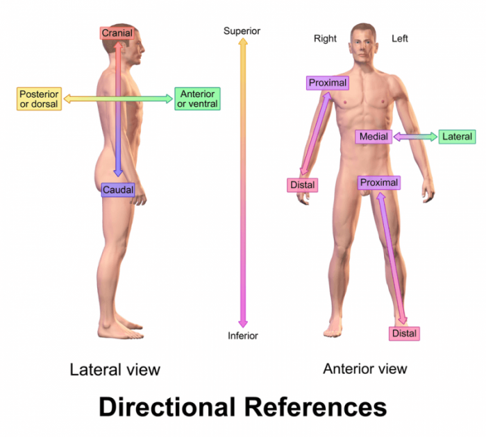
Photo: By National Cancer Institute – https://training.seer.cancer.gov/anatomy/body/terminology.html, Public Domain, https://commons.wikimedia.org/w/index.php?curid=2130068
Many structures in the body have names that include terms used to specify their position in the body relative to the anatomical planes or relative it to other structures in the body. Examples of these anatomical terms used to label body structures include the axial skeleton, the median cerebral artery, the posterior and anterior pituitary, and the inferior and superior vena cava.
Prefixes and suffixes are also used to indicate the position of anatomical structures within the body. The parathyroid glands have the prefix Para attached to them, referencing the fact that they are found on the thyroid’s posterior side. Similarly, the epidermis is the top layer of the skin, and the prefix “epi-” means outermost or uppermost. The adrenal glands are positioned at the top of the kidneys, and the “ad-” prefix means toward or near.
In addition to the terms for planes of the body, there are various anatomical terms used to give more precise descriptions of the location of a body structure. These are some of the terms created to assist in comparing the location of one body structure to the location of another.
The terms inferior and superior (or sometimes caudal and cranial) are used to denote whether a body part is facing the feet or head of the body. Inferior or caudal structures are those which face towards the feet, while superior or cranial structures of those which face towards the head. As an example, the inferior vena cava carries blood from the lower portion of the body towards the heart while the superior vena cava carries blood away from the head. Instead of inferior and superior the terms posterior and anterior are occasionally used. Though inferior and superior are the more common terms, posterior and anterior are occasionally used for interior body structures such as with the pituitary gland that has a posterior and anterior side.
On occasion, the ventral and dorsal are used to stand in for posterior and anterior, though typically they only wind up describing the anatomy of animals and not humans. The dorsal term refers to the upper or back side of an animal while the ventral term describes the lower or frontal side of the animal.
The term medial refers to structures that are closer to the middle of the body than they are the sides of the body, when viewing the body upright in the typical standing position. By contrast, lateral describes structures which are more towards the sides of the body. Many parts of the human body, like the lungs are considered symmetrical or bilateral within the central portion of the body.
Superficial structures are those that are closer to the surface or exterior of the body, as opposed to the body structures which are closer to the center of the body, the interior portion. Structures which are deeper in the body are, somewhat unsurprisingly, referred to as deep structures. Distal and proximal are two terms that are used to describe points in the body relative to the other point. For instance, a distal point refers to a point in the body farther away than the proximal point, which is closer. For instance, the proximal end of a leg is the end that joins it to the rest of the body, while the distal end is the far end of the extremity away from the center of the body.
Regional Terms
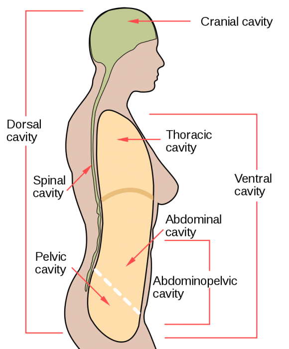
Photo: By NCI (original) / Mysid (SVG) – Vectorized by Mysid in Inkscape, based on . Image renamed from File:Body cavities.svg, Public Domain, https://commons.wikimedia.org/w/index.php?curid=5678872
Regional terms are used to divide different portions of the body into discrete regions. The appendicular region and the axial region are both different regions of the body. Appendicular is a term which refers to the appendages and limbs, structures which are connected to the main portion of the body. Meanwhile, the axial region refers to the primary portion of the body – the trunk, chest, neck, and head.
While those are the two primary terms, other regional terms referred to subsections of these two primary regions. As an example, the abdominal region is made out of a smaller portion of the axial region – the abdomen. In addition, the arms are part of the brachial region, a subsection of the appendicular region. The abdominal region can be divided into many different smaller regions composed of the various groups of tissues and organs.
Body Cavities
Body cavities are fluid-filled the spaces within complex, multicellular organisms. These body cavities are frequently regions/spaces where the internal organs of the body develop and take residence. The largest cavity within the human body is the ventral body cavity, which actually contains multiple cavities (the thoracic and abdominal-pelvic body cavities).
The cavities of the body include the dorsal cavity, the cranial cavity, the ventral cavity, the vertebral cavity, the thoracic cavity, and the abdominopelvic cavity. The dorsal cavity is one long continuous cavity that houses portions of the central nervous system including the spinal cord and brain. It is found on the body’s dorsal side. The cranial cavity contains the cerebrospinal fluid, the brain’s meninges, and the brain itself. The cranial cavity overlaps with the anterior portion of the dorsal cavity. The ventral cavity is where many different organs, such as the lungs, heart, liver, and spleen, can be found. Viscera is the term used to describe the organs found within the ventral cavity. The ventral cavity is divided by the diaphragm, a long stretch of skeletal muscles located beneath the lungs, with the two different regions being called the superior and anterior regions.
Body Quadrants
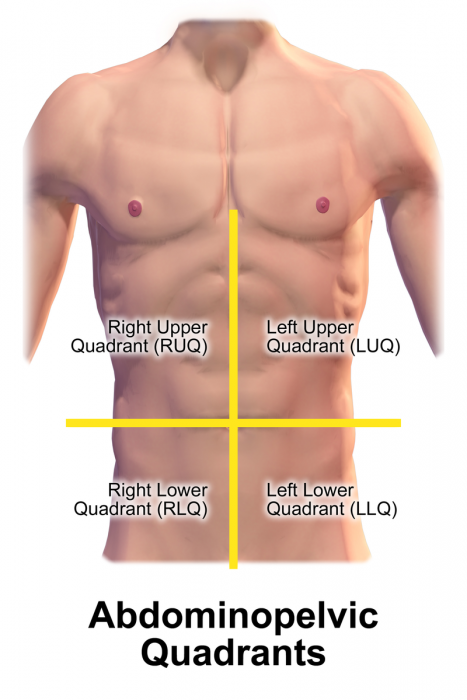
Photo: By BruceBlaus. When using this image in external sources it can be cited as:Blausen.com staff (2014). “Medical gallery of Blausen Medical 2014”. WikiJournal of Medicine 1 (2). DOI:10.15347/wjm/2014.010. ISSN 2002-4436. – Own work, CC BY 3.0, https://commons.wikimedia.org/w/index.php?curid=31339190
The abdominal section of the body can be divided into four different quadrants. These quadrants are defined by where the umbilical plane and the sagittal plane intersect. The left upper quadrant is where part of the stomach, the pancreas, the liver, the spleen, and the left kidney can be found. The right upper quadrant of the abdomen contains the right kidney, the gallbladder, the liver, the duodenum, the head of the pancreas, and sections of the small intestine.
The right lower quadrant of the abdomen is home to the appendix and the cecum. The right lower quadrant also contains the right half of the reproductive system for females and the right ureter. Finally, the lower left quadrant is home to portions of the large intestine, most of the small intestine, the left ureter, and the left region of the female reproductive system.
Related Posts
 Important Strategies To Help Healthcare Providers Support Patients With Diabetes
 Studying The Link Between Increased BMI And Late-Onset Preeclampsia In Pregnant Women
 A Bacterial Cell Imaging Method Using CRISPR And Microfluidics
 B. infantis Reduces Key Markers Of Intestinal Inflammation In Infants
 Epigenetic Changes In Multiple Sclerosis – Studied In Twins
 Did You Know Math Can Help Us Learn How Diseases Work?