


The anatomical human heart is the organ responsible for cycling blood throughout the human body, providing the tissues in the body with the oxygen they need. The heart is an integral part of the circulatory system, along with the blood vessels, blood, lymph and lymphatic vessels.
The heart works to circulate blood throughout the body, ensuring that the oxygen-rich blood carries oxygen to the tissues and organs that need it and that carbon dioxide and other wastes are removed from the blood. The heart’s anatomy is the result of evolutionary processes and necessary for its functions to be carried out properly.
The tissues within the human body require a constant delivery of oxygen and nutrition in order to stay alive and function. Should the heart cease pumping blood to the rest of the organs and tissues in the body, the body will die.
The Anatomy Of The Heart
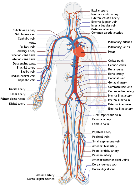
Rendering of the circulatory system. Photo: LadyofHats via Wikimedia Commons, Public Domain
The human heart is an organ roughly about the size of a fist and weighs between 8 to 10 ounces in women and 10 to 12 ounces in men. The anatomy of the heart is structured in such a way that it is said to have both plumbing and electricity. The heart is divided into four different chambers: the left and right atriums and left and right ventricles. The atriums are on top of the heart while the ventricles are on the bottom of the heart. The right atrium and right ventricle are said to comprise the “right heart” while the left atrium and left ventricle comprise the “left heart”. While the heart functions as a single organ, it is divided in the middle by a wall made out of muscle tissue. This muscle is called the septum.
There are also four different valves responsible for regulating blood flow through the heart. The function of the valves is to ensure that blood only flows in one direction, and that backflow doesn’t occur. The pulmonary valve is what controls how blood moves from the right ventricle over to the pulmonary arteries, while the tricuspid valve does the same for the right atrium and the right ventricle. The mitral valve lets the oxygenated blood coming from the lungs pass from the left atrium into the left ventricle. Finally, the aortic valve lets the oxygen-rich blood move from the left ventricle to the aorta.
The contractions of the heart are controlled by a system of electrical impulses which start in the sinoatrial node, which is found at the top of the heart’s right atrium. The sinoatrial node is what sends electrical impulses to the heart, which makes the myocardium contract. While the electrical impulses are sent at a constant rate, things like hormonal factors, stress, and physical needs may affect one’s heart rate.
The heart itself is encased by a double-walled structure referred to as the pericardium. This both provides the heart with protection and keeps it anchored at a specific place within the chest. The parietal pericardium is the outer layer of the pericardium itself, and the inner layer of the pericardium is dubbed the serous pericardium. In between the two layers is a fluid called pericardial fluid, which supplies the heart with lubrication during the contraction of the heart and the accompanying movement of the diaphragm and lungs.
The outer wall of the heart is made out of three distinct layers. The epicardium is both the outermost layer of the heart’s outer wall and the inner wall of the pericardium. The middle layer of the outer wall is the myocardium, and the muscles here is what causes the heart to contract. The inner layer of the outer wall is called the endocardium and it contacts the blood.
The heart has structures referred to as the atrioventricular valves, the function of which is to connect the ventricles and the atria. The mitral valve combined with the tricuspid valve form this valve group. There’s also a structure called the pulmonary semilunar valve that separates the pulmonary artery and the right ventricle. Similarly, the artic valve divides the aorta and the left ventricle. The chordae tendinae, or hearstrings, are what bind the valves to the heart muscles. The contraction of the heart is driven by electrical impulses created by a structure known as the sinoatrial node.
Supplying Oxygen And Pumping Blood
As previously mentioned, the function of the heart is to circulate blood throughout the body, getting rid of carbon dioxide and supplying the body tissue with oxygen. The heart accomplishes this task by circulating blood with two distinct systems: the systemic circuit and the pulmonary circuit.
The pulmonary circuit is responsible for taking deoxygenated blood and oxygenating it. The deoxygenated blood leaves the ventricle of the heart through the pulmonary artery, where it is carried to the lungs and oxygenated. The freshly oxygenated blood then uses pulmonary vein to return to the left atrium of the heart. Meanwhile, the systemic circuit is responsible for enabling oxygenated blood to reach the tissue of the body. In the systemic circuit, the oxygenated blood goes to the tissues of the body via the capillaries and arteries and supplies them with oxygen. The deoxygenated blood will then return to the heart using veins, heading to the venae cavae and entering the heart’s right atrium.
Given that the heart is a muscle as well, it also needs oxygen. How does the heart get its own supply of oxygen, so that it can continue to function? The heart gets its oxygen after the blood leaves the heart via the aortic valve. There are actually two sets of arteries that make sure some oxygenated blood gets to the heart. The left main coronary artery branches out and becomes the left circumflex artery and the left main coronary artery. The right coronary artery also branches out, but on the right side of the aorta, not the left.
Because these arteries supply the heart with oxygen so it can do its job, if these arteries become blocked they can lead to a heart attack. A cardiac arrest episode can potentially be different from a heart attack. Cardiac arrest is when the heart suddenly stops functioning correctly. This often occurs as the result of a disturbance of the heart’s rhythm, but other things may lead to cardiac arrest. Heart attacks may cause these problems but they aren’t the only way cardiac arrest can happen. Hears have their own natural pacemaker cells, which ensure that the heart contracts at the correct rhythm. The pacemaker cells all have the ability to set the rhythm or to follow the rhythm that has already been set. Those who medical issues like atrial fibrillation or irregular heartbeat have a problem where every cell is trying to set the rhythm of contraction, which means the cells beat out of sync.
Normal, healthy, heart contractions occur in five different phases. In the beginning phase – early diastole – the heart starts out relaxed. In the second phase, atrial systole, the atrium contracts and pushes blood into the ventricles of the heart. In the third phase, the ventricles begin contracting, though their volume doesn’t change. The fourth phase sees the ventricles contract while empty, and in the final phase the ventricles relax and cease contracting.
Related Posts
 Important Strategies To Help Healthcare Providers Support Patients With Diabetes
 Studying The Link Between Increased BMI And Late-Onset Preeclampsia In Pregnant Women
 A Bacterial Cell Imaging Method Using CRISPR And Microfluidics
 B. infantis Reduces Key Markers Of Intestinal Inflammation In Infants
 Epigenetic Changes In Multiple Sclerosis – Studied In Twins
 Did You Know Math Can Help Us Learn How Diseases Work?