


An octopus arm is one of the most fascinating organs among animals. It has many particular features enabling it to carry out various vital tasks like moving, touching, grasping, sucking, hiding and camouflage, mating, and smelling.
Among these features, a massive tridimensional array of muscle provides the arm with an almost unlimited “degree of freedom” for motion. The epithelia of its skin and especially that of its suckers are covered with a plethora of various sensory receptors (touch, taste,…) monitoring the animal environment.
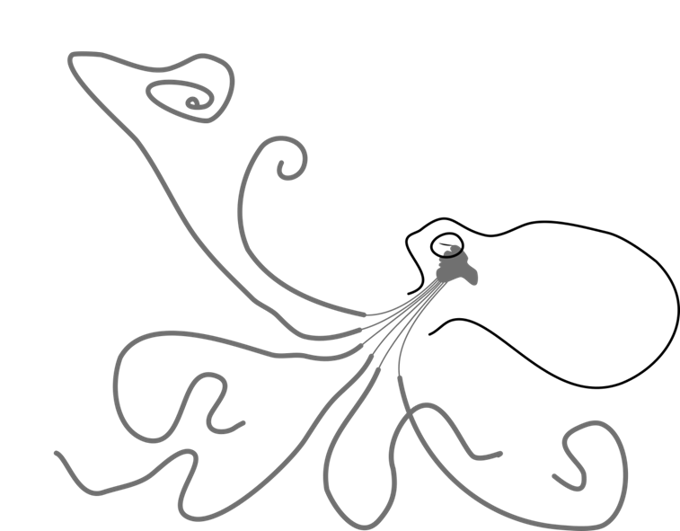
Most of all, each arm possess their own nervous system (Figure 1), and together they have more neurons than the central brain: the arms contain two third of the 500 million neurons that make an octopus nervous system (it is only a few hundred times less than a human brain that contains 86 billion neurons).
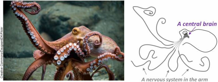
Figure 1: The octopus nervous system has a unique organization; it is divided into two components, one in his head, the central brain, and another in its arm. They contain about 170 and 350 million neurons, respectively. Credits:DaugaardDK/CC BY-NC-SA andJPB/CC BY-SA
A mesmerizing aspect of the arm nervous system is that it is not limited to process reflex or to act as a relay between the brain and the peripheral organs, but it shows a certain degree of autonomy (self-governance). The arm is able to process sensory information then organizes and executes a rudimental motor action by “himself”, although the central brain seems to keep control of the overall activity.
The central brain may ask: “grasp that crab” and the arm will “think” how to catch it.
In order to understand the basis of the neural processes taking place in the arm, we are gathering information on the chemical neuroanatomy of the arm nervous system, in particular, its neurotransmitters network. In other words, we try to understand: which neurons used which neurotransmitter, to communicate with which structure, and for which purpose. Since neurotransmitters are biochemically associated with particular psychological and physiological functions, we hope that studying their network might help to get a little closer to understand how “think” an octopus arm.
In a recent work, we focused on the identification and visualization of the monoamine neurotransmitter 5-hydroxytryptamine (better known as “serotonin”) in the octopus arm. In vertebrates, serotonin is implicated, for instance, in cognition, mood, anxiety, aggression, sleep or appetite, but it is also involved, in the modulation of the sensory mechanism of neurotransmission. Because neurons synthesizing serotonin cannot be observed directly in tissue, therefore we applied an immunohistochemical method for imaging serotonin molecules. This is a method using “hacked” antibodies made to recognize a specific molecule (in the present case specifically serotonin, no other monoamines), and modified with a label observable using a light microscope. Applying this method, we were able to visualize the neurons synthesizing serotonin, and generate for the first time a schematic map of serotonin innervation in the octopus arm.
Our observations revealed two types of serotoninergic innervation. One type, which arises from the brain, innervates only the periphery (skin, blood vessels, or both) and comprises a few serotoninergic efferent fibers running in the cerebrobrachial tract (a kind of super-highway of nerve fibers at the center of the arm connecting the brain and the periphery). Another type originates the neurons (Figure 2) localized in the axial nerve cord (the main nervous center that runs at the center of the arm from its base to its tip.), and forms an intrinsic network in the arm by projecting notably in direction of (i) the intramuscular nerve cords (four small nerve cords also running all along the arm in a quadrant which the center is the axial nerve cord); (ii) the ganglia that innervated each sucker; (iii) the sucker. Interestingly these innervation seems to preferentially terminate close to structure with sensory function (such as the sensory cells of the sucker epithelium), suggesting that serotonin might play a modulatory role in the sensory pathway of the octopus arm.
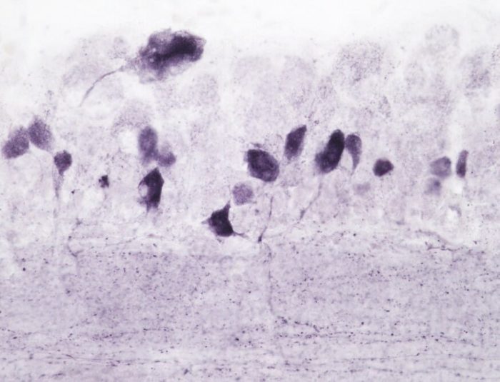
Figure 2: Neurons synthesizing the neurotransmitter serotonin in the octopus arm are labeled in purple. Credits: by JPB/CC BY-SA
In 1908, Guerin using basic histological methods described for the first time neuroanatomical organization of an octopus arm, a work completed thereafter by researchers such as May and Martoja, Graziadei or Young. Using specific imaging methods, we were able to visualize serotonin and provide a detailed map of its innervation in the octopus arm. Our observation revealed an intrinsic serotoninergic innervation that may participate in the modulation of the sensory transmission and brings new neuroanatomical evidence supporting the concept that an octopus arm is somehow autonomic.
[Note from the authors: While out of the scope of this synopsis, the authors wonder how the central brain manages its relationship with its eight arms. Does the central brain ask its arms: “grasp that crab” or “Please, grasp that crab”? Does the octopus central brain sometimes wonders: “Where is my mind?”
These findings are described in the article entitled Immunohistochemical and biochemical evidence for the presence of serotonin-containing neurons and nerve fibers in the octopus arm, published in the journal Brain Structure and Function. This work was led by Jean-Pierre Bellier, Yu Xie, Sameh Mohamed Farouk, Yuko Sakaue, Ikuo Tooyama, and Hiroshi Kimura, from the Shiga University of Medical Science and Beihua University.
Related Posts
 Improving Tools For Quality Improvement: Crossings, Runs, And Crossrun
 Coherent Poly Propagation Materials With 3-Dimensional Photonic Control Over Visible Light
 Reconstructing Commuter Networks Using Machine Learning And Urban Indicators
 Surprising Internal Structure Of Whole Wheat-Green Gram Functional Bread
 Tailoring Tomatoes To Match Individual Consumer Needs
 Eavesdropping On “Classroom Talk” In Undergraduate STEM Classrooms