


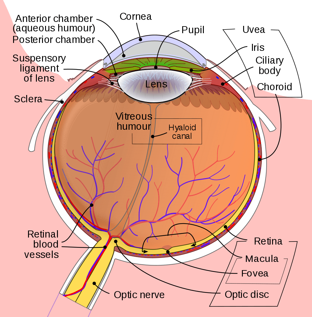
The human eye is composed of many different parts that work together to interpret the world around us. What you want to interpret as a major part of the human eye is somewhat up to the individual, but in general there are seven parts of the human eye: the cornea, the pupil, the iris, the lens, the vitreous humor, the retina, and the sclera.
Let’s take a closer look at each of these components individually.
The Cornea
The cornea is the outermost portion of the eyeball. The responsibility of the cornea is to focus the light that enters our eyes. The cornea is transparent, and it covers the pupil, iris, and anterior chamber. The cornea itself is composed of five different layers, and the function of the outermost layer is to protect the eye from dirt and foreign objects, augmented by tears which keep the eye moist and clean out dirt.
The cornea is one of the most sensitive regions of tissue in the entire body, owing to the fact that it has many, densely connected nerve fibers running through it. The receptors in the cornea could have concentrations somewhere between 300 to 600 times greater than the density of the pain receptors in a patch of skin.
The cornea of the eye is composed of five different layers: the corneal epithelium, Bowman’s layer, the corneal stroma, Descemet’s membrane, and the corneal endothelium. Each of these layers has a function and they work together to transform the light entering the eye as well as protect and support the eye in general.
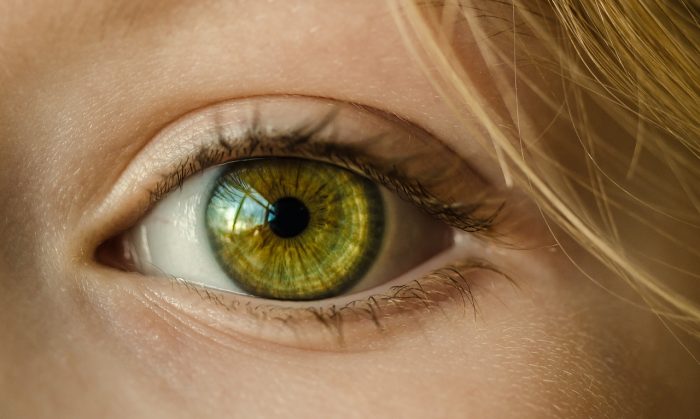
Photo: (https://pixabay.com/photos/eye-iris-look-focus-green-1132531/) Skitterphoto via Pixabay, Pixabay License (https://pixabay.com/service/license/)
The Iris
The iris is a structure found in the eyes of most mammals, and along with the pupil it controls how much light enters the eye. The iris is comprised of two different layers: the stroma, and the pigmented epithelial cells. The upper layer, the stroma, is linked to muscles that contract and dilate the pupil. The iris is divided into six different layers: the anterior layer, the stroma of iris, the iris sphincter muscle, the iris dilator muscles, the anterior pigment epithelium, and the posterior pigment epithelium. The two muscles found in the eye are what control the dilating and contraction of the pupil.
The rear portion of the iris is filled in with pigmented epithelial cells. The function of the pigment is to prevent light from penetrating the retina, ensuring that the only region where light enters the eye at is the pupil. The pigment of the eye is typically brown, gray, green, hazel, or blue in coloration. Although there are some rare conditions that can make the iris colors like a pinkish-white.
The Pupil
The pupil isn’t really a structure in the traditional sense, although it is still a major component of the eye. There pupil is an opening within the iris that allows light to enter the eye, channeling the light toward the retina of the eye, where it will be collected and converted into electrical signals. The pupil of the eye is usually almost perfectly round and black, and the dark black appearance of the pupil results from the fact that the pupil isn’t reflecting any light back, rather it is absorbing the light. The pupil and the iris work together to control the amount of light that enters the eye. If you want to think about it in terms of a camera lens, the eye’s aperture is the pupil and the iris controls the aperture.
The size of the pupil is affected by small muscles found in the iris. One muscle is responsible for dilating the pupil, while the other muscle is responsible for constricting the pupil. The pupil dilates in conditions of low light to improve vision, while in bright conditions the pupil will constrict, imposing a limit on how much light can enter the eye. The size of a person’s pupil can change with age, with children having larger pupils than adults. A typical adult has a pupil size of roughly 2 to 4 mm undilated and 4 to 8 mm when dilated in the dark.
The Lens
The lens of the eye, much like a camera lens, is what changes the eye’s focal distance, bringing things in and out of focus. The lens of the eye takes the light that has passed through the pupil and bends them in ways that allow the retina to collect clear images of objects. These objects can be located at many different distances, but the lens is able to focus on them all. The lens focuses light with the assistance of the cornea.
The lens is in a biconvex, ellipsoid shape. Biconvex means that the lens is rounded on both halves, or sides, and the fact that its an ellipsoid means that it is a stretched curve, like the shape of an olive. The lens changes shape as it shifts its focus, but in general, the lens is about 4 mm in width front to back and 10 mm across in adults.
The lens is made out of three different parts: the lens fibers, the lens epithelium and the lens capsule. The lens capsule is the outermost layer, and it is a smooth transparent layer that fits over the lens epithelium and fibers. The lens epithelium is found under the capsule and it has the responsibility of stabilizing the lens and creating lens fibers. The lens fibers are the innermost layer of the lens and they are thin, long, transparent fibers that compose most of the lens’ volume.

Photo: (https://commons.wikimedia.org/w/index.php?curid=29025014) By BruceBlaus. When using this image in external sources it can be cited as:Blausen.com staff (2014). “Medical gallery of Blausen Medical 2014”. WikiJournal of Medicine 1 (2). DOI:10.15347/wjm/2014.010. ISSN 2002-4436. – Own work, CC BY 3.0, (https://creativecommons.org/licenses/by/3.0)
The Vitreous Humor
The vitreous humor is comprised of a clear substance that fills in the area between the back of the eye and the retina. The eye has to process visual data, and because of this the gel-like vitreous humor must be clear enough that light can filter through it. The vitreous humor is an immobile fluid, and it isn’t replenished or regenerated in any way, it is also not served by blood vessels. Substances which enter the humor will stay floating in the gel of the humor, and this causes “floaters” which can interfere with a person’s vision. The vitreous humor tends to thin as people age.
The Retina
The retina is responsible for converting light into electrical signals that will be carried to the brain for processing. The retina uses photoreceptor cells to process the light that comes through the vitreous. The light-sensitive cells pick up both light-intensity and color and transmit these to the brain. The layers of the retina are comprised of neurons joined together by synapses. Two different types of cells capture the vast majority of the light that hits the retina: cones and rods. Cones are cells responsible for interpreting color, and they function only in well-lit environments. In contrast, rods interpret black and white vision, and they function in conditions of dim light.
There are two distinct layers of the retina. These layers are responsible for four different stages of processing: photoreception, the transmission of the received info to bipolar cells, the transmission of the information to the ganglion cells, and transmission of the signal along the optic nerve.
The retina routes the signals it receives through the optic nerve, and when the brain receives these signals it interprets them. Damage to the retina can cause irreversible blindness, as the brain cannot receive visual stimuli.
The Sclera
The sclera us also referred to as the white of the eye, and it is comprised primarily of collagen and elastic fibers. The sclera is an opaque substance that functions to support and protect the rest of the eye, helping maintain the globe shape of the eye and resist both external and internal forces. It is made out of four layers: the episclera, the stroma, the lamina fusca, and the endothelium.
While some animals have a pupil large enough to obscure most of the sclera, humans have pupils that are relatively small in comparison and where the sclera is prominently shown. Because of the fact that this makes determining where an individual is looking simpler, it is possible that the size of the pupil and the sclera evolved as a method of nonverbal communication.
The Occipital Lobe
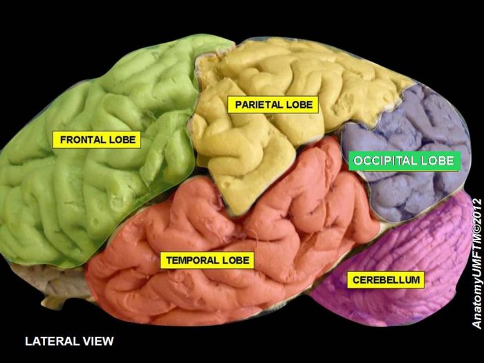
Photo: (https://commons.wikimedia.org/w/index.php?curid=19131987) By Anatomist90 – Own work, CC BY-SA 3.0, (https://creativecommons.org/licenses/by-sa/3.0)
While not part of the eye, the occipital lobe plays an incredibly important role in our vision. The occipital lobe is found at the back of the brain, and it is the visual processing center of the human brain. The occipital lobe it takes the electrical signals originating from the retina and carried via the optic nerve, and converts these signals into our visual perception of the world. If the occipital love sustains damage, partial or complete blindness can end up happening. The occipital lobe is divided into several different regions, including the primary visual cortex, which is itself subdivided into the dorsal stream, the ventral stream, and the dorsal medial area.
Related Posts
 Important Strategies To Help Healthcare Providers Support Patients With Diabetes
 Studying The Link Between Increased BMI And Late-Onset Preeclampsia In Pregnant Women
 A Bacterial Cell Imaging Method Using CRISPR And Microfluidics
 B. infantis Reduces Key Markers Of Intestinal Inflammation In Infants
 Epigenetic Changes In Multiple Sclerosis – Studied In Twins
 Did You Know Math Can Help Us Learn How Diseases Work?