


The brain is a fascinatingly complex organ, responsible for all that we do and experience. The brain is so complex that even to simply discuss it, certain distinctions have to be made regarding its structure, and for that reason, scientists divide the brain up into three major portions with each of these portions divided into many subregions and structures. The main portions of the brain are the cerebrum, the cerebellum, and the brainstem.
Let’s take a detailed look at the three major portions of the brain, exploring their general structure and function.
Cerebrum
The cerebrum is the largest and most complex portion of the human brain. The cerebrum’s function is to control our actions and thoughts, either conscious or unconscious, and responses to stimuli. The cerebrum itself is typically divided into four different lobes: the temporal lobe, the parietal lobe, the occipital lobe, and the frontal lobe.

Photo: Surface of the cerebrum, (https://en.wikipedia.org/wiki/Cerebrum#/media/File:Lateral_surface_of_cerebral_cortex_-_gyri.png) by Patric Hagmann et. al. via Wikimedia Commons, CC BY 2.5 (https://creativecommons.org/licenses/by/2.5)
The temporal lobe of the cerebrum is responsible for the perception of auditory information, and it handles both memory and speech functions. The hippocampus is located within the temporal lobe and its function is to handle the formation of new memories. The hippocampus also plays a role in the association of memories with emotions, and the ability to learn associations between concepts. If this region of the brain is damaged, problems with the formation of language and memories will manifest. The pituitary gland is found in the temporal lobe as well, and it is linked to the hypothalamus through a structure known as the pituitary stalk. The function of the pituitary gland is to manage levels of hormones within the body, specifically those that control and stimulate sexual development, handle stress responses, encourage the growth of bones and muscle, and fight off disease. The pituitary gland accomplishes this by controlling other glands within the body, the endocrine glands.
The parietal lobe is located on top of the brain, and its primary functions include the perception of stimuli as well as the recognition of objects, movement, and orientation. The parietal lobe analyzes visual motion, controls our attention, orients us in space, and analyzes the space around us by interpreting the signals we received from our senses. In addition, regions of the parietal lobe are important to the processing of language.
The occipital lobe is found at the back of the brain. The occipital lobe is mainly responsible for the processing of visual information. The primary visual cortex is a structure located within the occipital lobe, and it receives information collected by the retinas. The occipital lobe than interprets these signals and gives us our experience of vision. Damage to the occipital lobe will result in visual impairments/confusion.
The frontal lobe of the brain is responsible for our critical thinking, planning, reasoning, and problem-solving, as well as our experience of emotions. The rear portion of the frontal lobe is the motor cortex, which receives inputs from the other lobes and carries out the movements of the body associated with them.
The structure that results from the joining of these four regions is referred to as the “cerebral cortex”, which can itself be divided into different halves. The two halves of the cerebral cortex are the left and right hemisphere, and it is often claimed that the right hemisphere handles more creative tasks while the left hemisphere handles tasks correlated with reason and logic. However, this model oversimplifies things somewhat, as both halves/hemispheres are needed to carry out both creative and logical actions.
Within the cerebrum is a structure referred to as the neocortex, which is comprised of six different layers. The neocortex is only found within mammals, and research on the structure suggests that it is a fairly recent product of evolution. It could be one of the factors responsible for the sophisticated information processing witnessed in humans, as well as other higher mammals like dolphins and other primates. In small animals like rats, the neocortex is fairly smooth, but in the higher mammals it is filled with large wrinkles and grooves called gyri and sulci respectively.
A structure called the corpus callosum is responsible for joining the two hemispheres of the cerebral cortex together, and it made out of a thick bundle of axon fibers, containing of 2000 million axons. The sheer number of these fibers makes communication between the two halves of the brain easy and the corpus callosum is also where the largest collection of white matter in the brain is found.
The white matter is the brain material responsible for carrying signals that originate in the nerve cells and head to other parts of the body and brain. You can think of the white matter as something that enables communication between different parts of the brain, while the gray matter is used for the purposes of memory and computation/thinking. The top portion of the brain is all gray matter, and if you were to look at the brain you would be seeing mainly gray matter.
Cerebellum
“Cerebellum” comes from Latin and it roughly translates to “little brain”. The cerebellum is part of the hindbrain in humans and all vertebrates. The cerebellum gets information from the sensory parts of the brain, as well as the spinal cord, and uses this information to regulate motor movement. The cerebellum functions to control our posture and balance, keep our muscle activity balanced, and coordinate our movements and speech. The cerebellum also plays a major role in the learning of motor behaviors.
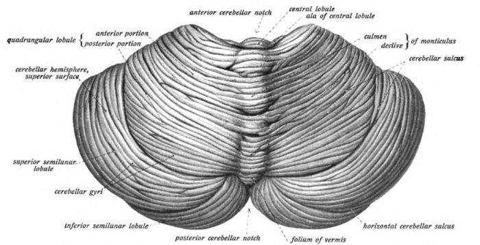
Crerebellum from behind/above (https://en.wikipedia.org/wiki/Cerebellum#/media/File:Sobo_1909_653.png) by Dr. Johannes Sobotta – Sobotta’s Textbook and Atlas of Human Anatomy 1908 via Wikimedia Commons, Public Domain
Anatomy wise, the cerebellum is located just behind the pons and medulla of the brainstem, near the fourth ventricle. The cerebellum can be divided into three discrete lobes: the anterior lobe, the posterior lobe and the flocculonodular lobe.
The flocculonodular lobe Is the oldest part of the cerebellum in terms of evolution , and this is the structure responsible for spatial orientation and balance. The medial region of the posterior and anterior lobes function to control fine body movements, taking in input from the spinal cord as well as the auditory and visual systems of the brain. The lateral region of the cerebellum is the largest part of the cerebellum in humans. This region gets inputs from the cerebral cortex. It’s suspected that it plays a role in the planning of movements which are going to occur shortly, and in the evaluation of sensory inputs required for action.
The cerebellum is referred to as the “little brain” due to its small size when compared to the cerebrum. In fact, the cerebellum only accounts for about ten percent of the brain’s total weight. Despite this, about half of the brain’s neurons are found within the cerebellum, and the neurons are responsible for transmitting information through electrical signals to other portions of the brain and body. While the cerebrum and neocortex are features fairly unique to higher mammals, the cerebellum is not. The cerebellum is an old brain structure and scientists believe that animals which existed long before human possessed the cerebellum.
If damage occurs to the cerebellum, although intellectual impairment and paralysis may not occur, fine motor problems like slower movement, unsteadiness, and tremors may manifest. It would be difficult to carry out complex physical tasks under these conditions.
Brainstem
The brainstem is found at the very bottom of the brain, connecting the rest of the brain to the spinal column. It is the oldest region of the brain in terms of the brain’s evolution. The brainstem is responsible for the regulation of actions necessary for survival such as breathing, sleep, digestion, heart rate, and reflexes. The cranial nerves run through the brainstem and function as a control system for all the signals coming from the peripheral nervous system and heading towards the brain. Examples of the nervous system pathways that utilize the brainstem and cranial nerves include the spinothalamic tract and the corticospinal tract.
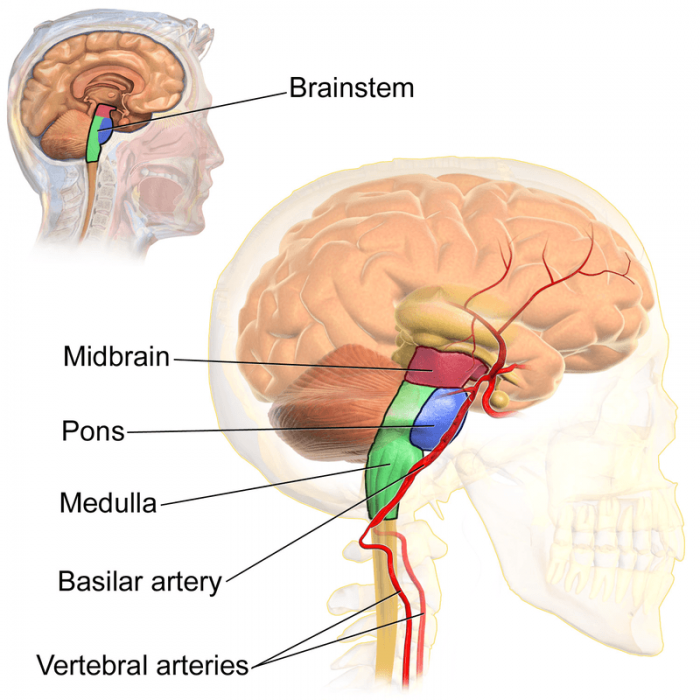
Photo: Vertebral arteries and the brainstem (https://en.wikipedia.org/wiki/Brainstem#/media/File:Blausen_0114_BrainstemAnatomy.png) by BruceBlaus via Wikimedia Commons, CC by 3.0 (https://creativecommons.org/licenses/by/3.0)
The brainstem is divided into three different regions by anatomists: the midbrain, the pons, and the medulla.
The midbrain, which is often referred to as the mesencephalon, is the part of the brainstem found highest in the brain. The midbrain connects the other portions of the brainstem to the cerebrum and cerebellum. The midbrain is divided into three different regions: of the tectum, the tegmentum, and the ventral tegmentum. Many nervous tracts in the body pass through the tegmentum, which functions as a relay point for the motor signals carried by these tracts.
The second region of the brainstem is the pons, and it is found in between the midbrain and the medulla. The pons is responsible for a variety of different functions, such as controlling your breathing while you are sleeping, swallowing, eye-movement, and facial sensation. The pons also functions as a relay center for signals traveling between the cerebrum and cerebellum.
The medulla oblongata, frequently just called the medulla, is the lowest portion of the brainstem, and it is also thought to be the oldest part of the brain. The medulla is responsible for the control of heart rate, sneezing, breathing, and other automatic functions of the body. The upper, thicker, region of the medulla is connected to the fourth ventricle, while the lower portion of the medulla is joined to the spinal column. The vast majority of the nerve signals in the body have to travel through the medulla when they head to the brain from the body, or vice versa.
Related Posts
 Important Strategies To Help Healthcare Providers Support Patients With Diabetes
 Studying The Link Between Increased BMI And Late-Onset Preeclampsia In Pregnant Women
 A Bacterial Cell Imaging Method Using CRISPR And Microfluidics
 B. infantis Reduces Key Markers Of Intestinal Inflammation In Infants
 Epigenetic Changes In Multiple Sclerosis – Studied In Twins
 Did You Know Math Can Help Us Learn How Diseases Work?