The study of bone microanatomy provides complementary knowledge which is unavailable in most of the morphological analysis of fossil vertebrates. New information about growth, metabolic rates, and lifestyle pop up after analyzing thin sections of tetrapod bones under microscopy.
Moreover, bone histology can also detect features that indicate degrees of bone specializations, such as osteosclerosis. It means that there is an increase in the inner bone compactness which can be related to semiaquatic reptiles and has been already found in the forelimb of a semiaquatic crocodile from Brazil. On the other hand, there is osteoporosis, a porous pattern linked to the deficiency in secondary bone deposits during remodeling and associated with a bone mass decrease in long and shell bones of fast-swimming animals.
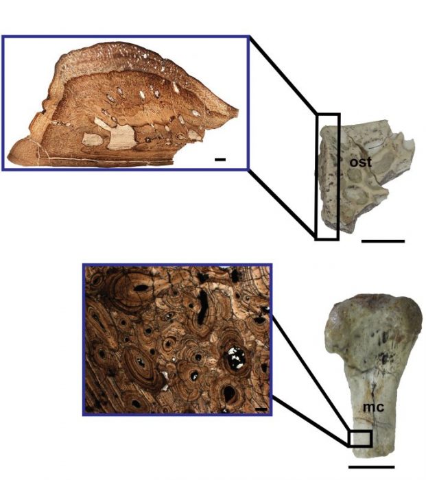
In our recent study on a bone microanatomy of Pepesuchus deiseae, published in the journal Cretaceous Research, we have sectioned and prepared thin sections of bones belonging to Pepesuchus deiseae, a Cretaceous crocodyliform which lived in Brazil during the Gondwana. In the Metacarpals, we observed the predominance of osteosclerosis organization. This specialization could be acting as a ballast to aid in buoyancy control during swimming. The same feature has been recorded in the ulna of Susisuchus anatoceps, a crocodyliform from Early Cretaceous. Also, the Haversian bone tissue was found by the presence of secondary osteons, resulting from bone remodeling.

Microanatomy of an osteoderm (ost) and a metacarpal (mc) of Pepesuchus deiseae. We can see several erosion cavities on the thin section of the osteoderm. Moreover, metacarpal’s thin section shows an extensive remodeling represented by Haversian bone. These features seem to belong to a adult female animal, which already accomplished some ovogenetic cicles.
Stéphane Hua and Vivian de Buffrénil published an article in 1996 reporting this same tissue in the humerus of a marine Jurassic crocodyliform, Steneosaurus. The authors suggested this bone remodeling pattern is linked to calcium metabolism for ovogenesis. Hence, this bone element might belong to a mature female. The dermic scute (osteoderm) of the Pepesuchus exhibits erosion cavities (Figure 2), which probably evidences bone reabsorption to egg formation. Early studies indicate that a crocodile’s osteoderms are a source of calcium storage, and the ions are mobilized from there during ovogenesis. Osteoderms present high microanatomical plasticity, and their arrangement changes according to the metabolic needs. Osteoderm and metacarpal micromorphology indicate the presence of pattern that can be related to mature females.
In addition, the External Fundamental System (EFS), or also named Outer Circumferential Lamellae (OCL), were found on the periosteal surface of the tibia confirm that this specimen had an advanced ontogeny development. These layers are deposited in the outermost cortex (periosteal region) of adult archosaurs (the group which includes dinosaurs, pterosaurs, and crocodiles) and mammals. This new data reveals that bone histology of fossil tetrapods may explain the processes occurring inside the animal before its death.
These findings are described in the article entitled Bone microanatomy of Pepesuchus deiseae (Mesoeucrocodylia, Peirosauridae) reveals a mature individual from the Upper Cretaceous of Brazil, recently published in the journal Cretaceous Research. This work was conducted by Mariana Sena, Rafael Andrade, and Juliana Sayão from the Universidade Federal de Pernambuco and Gustavo Oliveira from the Universidade Federal Rural de Pernambuco.


