


Unlike humans or other animal species, dolphins rarely show signs of external aging. Consequently, estimating the age of a free-ranging dolphin during a veterinary examination is particularly challenging. Previous methods to estimate dolphin age have relied on tooth extraction and counting the growth layer groups within the tooth to estimate age. We wanted to provide an alternative, less invasive, and, if possible, a more precise methodology which could accurately estimate age throughout the dolphin’s life span.
Dr. Daniel García-Párraga at Oceanogràfic in Valencia, Spain, created a novel methodology to estimate the age in dolphins via radiography of their pectoral flipper following similar principals applied to human hands. The collaboration between marine mammal veterinarians and human radiologists has been decisive in facilitating and orienting this work. We collaborated with human radiologist Dr. Luis Martí-Bonmatí and bioinformatics engineer Dr. Roberto Sanz-Requena to extrapolate from the human hand methodology and apply this to dolphin flippers.
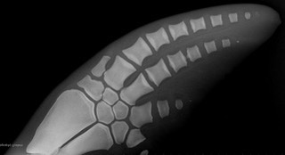
Figure provided courtesy of the authors
“There is so much potential in the collaboration between veterinarians and human physicians to further advance in the field of medicine. This is a clear example on how we can improve and better understand the dolphin life cycle just validating and adapting a technique that has been routinely used in the human medicine field for over 50 years,” Dr. García-Párraga explains.
The aim of this project is to facilitate accurate age estimation of free-ranging bottlenose dolphins during veterinary physical examinations, as part of conservation population health assessments. Also, for all animals of known age born under human care, this technique permits the detection of abnormal growth or developmental disorders, following the same approach that is routinely used as a diagnostic tool in children.
Conservation medicine veterinarian Dr. Ashley Barratclough at the National Marine Mammal Foundation worked with Dr. García-Párraga to finalize this project, which was recently published in Plos One.
“The aim is to use these methods in future health assessments of dolphins impacted by the Deepwater Horizon oil spill to interpret their biological data and understand the health issues experienced in this population. Accurate age estimation allows us to understand the reproductive capabilities of the individual and ultimately understand the population demographics and predicted survival,” Dr. Barratclough says.
For example, certain blood sample measurements would be expected to change in an older dolphin such as liver or kidney values; however, changes in these organs in a young dolphin would be of much greater concern to the veterinarians.
Over the last 15 years, a database of 126 radiographs has been compiled from multiple institutions in an unprecedented collaborative effort, in order to bring Dr. García-Párraga’s vision into practice. By analyzing each radiograph individually and assigning a score to each bone, we were able to determine the specific chronological changes which occur during each period of the dolphin’s life. As male and female dolphins grow at different rates, we had to make two separate formulas to ensure increased accuracy of age estimation. From their pectoral flipper radiographs, the dolphin’s age can be predicted within a few months in young dolphins (<5 years old) to within a few years if the animal is > 25 years old.
“This technique is an exciting advancement in the field of conservation medicine as it will enable an accurate less invasive method of estimating dolphin age and subsequently interpreting their veterinary physical exam accurately,” Dr. Barratclough says.
This novel diagnostic tool could also be applied to diagnose atypical ossification patterns consistent with nutritional, developmental, or growth abnormalities, and identifying subclinical health issues. The authors evidence, based on cases of fully-known history being under human care, that individuals suffering from severe illness during certain periods of their lives can have an impact on their bone ossification pattern.
This method could also be applied to stranded cetaceans to help to understand the reason for stranding and whether there is an age component to the underlying cause. The technique also permits aging stillbirths or abortions based on the level of ossification of the carpal region while still in the uterus. Even museum specimens in marine mammal collections could be aged without needing to destroy or manipulate bone or dental material.
Besides invasiveness, one important limitation when working with traditional techniques is that most marine mammal species are protected, and tissues can only be collected or transported under very strict permission. The benefit of different radiographs is that they can be easily distributed worldwide to the requested specialist or even interpreted immediately.
In addition, knowledge of the lifespan and the onset of sexual maturity for each species may allow this model to be applied to other cetaceans with similar bone maturation developmental curves, facilitating age estimation via pectoral radiography in future research.
Related Posts
 Duck-Billed Dinosaurs Uncovered In Aniakchak, Alaska
 Cryptic Diversity In Vietnam’s Limestone Karst Habitats
 An Improved Method To Remove Debris From Cyst Nematode Egg Suspensions And Computer-Aided Technologies For Egg Counting
 The Footprints Of Urbanization, Industrialization, And Agriculture On River Beds: Heavy Metal Contamination Assessment And Source Identification In River Sediments In Eastern China
 Glycoalkaloids In Potatoes: The Effect Of Biostimulants And Herbicides
 Tracking Cats: Using Stable Isotopes To Geographically Place Mammalian Carnivores