


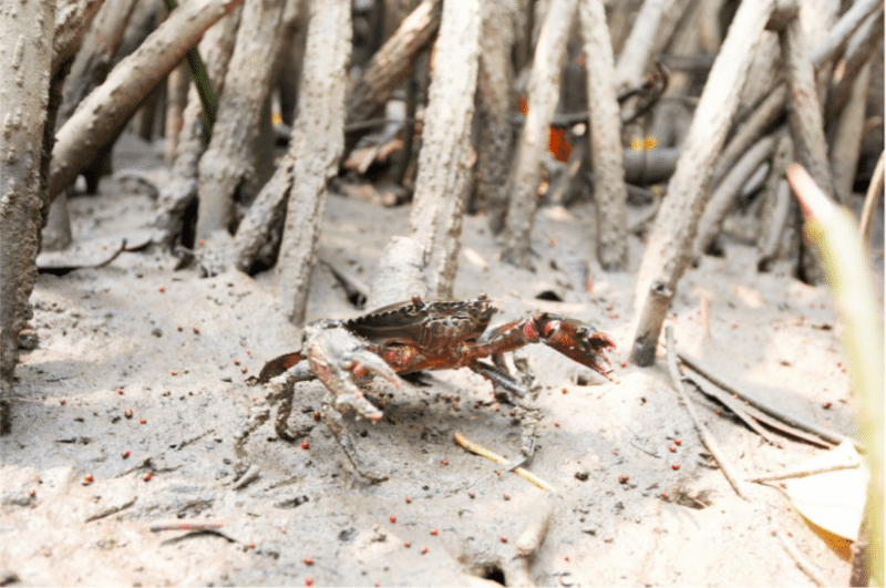
On the outside, they look exactly like the normal mud crabs that we loved to eat so much; Nothing much has changed, at least not at first glance. However, some of these crabs have been parasitized by microscopic barnacles known as sacculinids. Upon infection, they bend the infected host crabs to their will, feminize male crabs, castrating infected hosts, and ultimately, the hosts become living incubators for more sacculinid babies. The infected hosts still perform normal routines including feeding, but they are not able to reproduce anymore, thus the nickname “zombie crabs.”
The life cycle of sacculinids is straightforward: almost everything revolves around the host. The female sacculinid first infects the host by settling on its external cuticle or gill filaments. Once settled, it will invade the haemolymph (blood system) of the host, slowly forming root system (interna) to absorb nutrient. Once ready, the sacculinid will produce an external sac known as externa on the crab’s external abdomen, and within it, contains thousands of eggs. Fertilization occurs when a male sacculinid enters the externa, and once that happens, myriads of fertilized eggs will be released into the water, ready to infect new crabs. Therefore, if the population is dense, it only takes a few infected crabs to infect the whole population.
Such infection of sacculinids (Sacculina beauforti) on mud crabs (Scylla olivacea) is found to occur at Marudu Bay, Sabah, Malaysia. The high infection rate, with about 42% of the screened mud crabs S. olivacea infected, is alarming, especially when this mud crab species is highly sought-after for human consumption purposes. To make matter worse, we found that most of these infected crabs were sold off at a higher price as berried females (females carrying eggs) in the local market. The effect on humans after consuming these infected crabs, along with the parasites, is still unknown, but at least we know now that the external sac on the crab’s abdomen is not crab eggs but instead, the egg sac of a parasite.
Although the presence of an externa is the most commonly used indicator of sacculinid infection, this feature is not always visible as some crabs were reported to show altered morphological characters but with no externa or scar of lost externa. Aside from changing the host’s behavior, major changes are also observed in the host’s external morphologies. Among some of the altered characters include the widened abdomen and shortened gonopods of the males and shortened pleopods of the females (Figure 1 & 2).
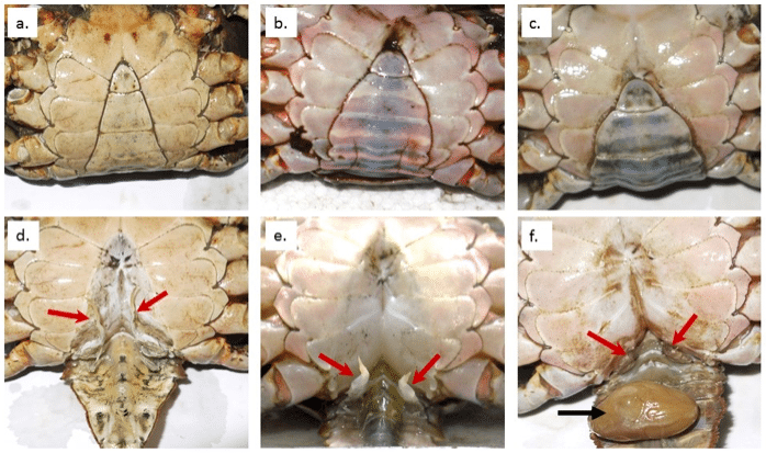
Figure 1. The abdomen morphology and gonopod (red arrows) of normal male (a & d), infected male without externa (b & e) and infected male with externa (c & f). Externa is marked with a black arrow. Figure 1 and 2 are adapted from Fazhan et al., 2018.
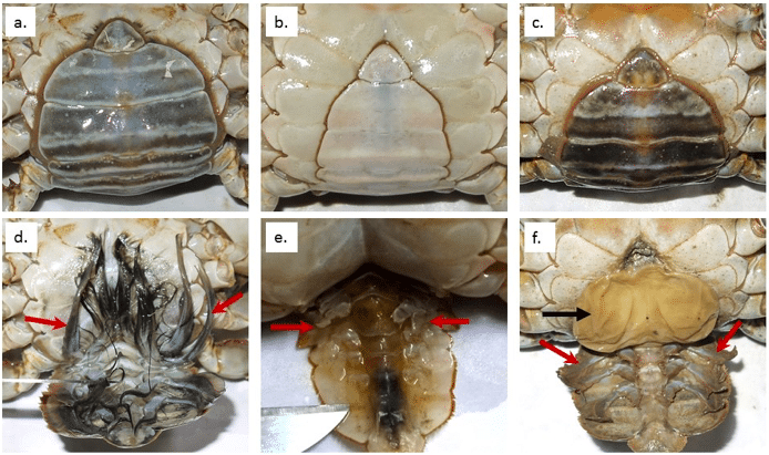
Figure 2. The abdomen morphology and pleopod (red arrows) of normal female (a & d), infected female without externa (b & e) and infected female with externa (c & f). Externa is marked with a black arrow. Figure 1 and 2 are adapted from Fazhan et al., 2018.
To ease their identification, especially when externa is not visible, we documented the morphological changes that are obvious in all infected crabs and constructed two formulas based on stepwise discriminant function analysis, allowing a better prediction of the infection status when no externa could be found. Function 1 and 2 are only applicable in males and females, respectively:
![]()
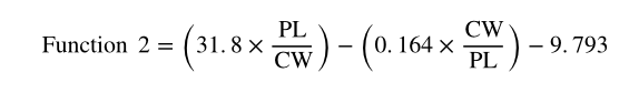
Where AW = abdomen width, CW = carapace width, GL = gonopod length, PL = pleopod length.
The cut-off values for male is -0.09 (≤-0.09 = infected males) and for female is -1.76 (≤-1.76 = infected females). These two equations are highly accurate, with a high success reclassification rate of 100% and 94% in the infected females and males, respectively. To further test the power of the constructed equations, both functions were fitted with data of normal crabs from three geographically distinct locations. A total of 100% and 95% classification success in females and males were recorded. This proves the feasibility of the constructed functions using discriminant function analysis to distinguish between normal and infected S. olivacea. This method is simple, non-invasive to the inspected crabs and can be performed by anyone without the requirement of specialized skills. The only two equipment needed are Vernier caliper and a calculator.
Stepwise discriminant function analysis has been used in species identification and sex determination of species with size dimorphism, such as birds. Our study represents the first successful attempt to predict sacculinid infection status of a commercially important crab species using such analysis. By only having to measure four variables (AW, CW, GL, and PL), the two proposed functions allow fishermen and farmers to select only healthy crabs for sale and re-stocking purposes. This will not only ensure that the infected crab population is contained but also guarantee that only healthy crabs are to be sold to end consumers.
These findings are described in the article entitled Predicting the sacculinid Sacculina beauforti infection status of the orange mud crab Scylla olivacea by discriminant analysis, recently published in the journal Aquaculture. This work was conducted by Hanafiah Fazhan, Khor Waiho, and Hongyu Ma from Shantou University, Mhd Ikhwanuddin and Mohd Ago Surzanne from the Institute of Tropical Aquaculture, Universiti Malaysia Terengganu, and Hin Boo Wee from the University of the Ryukyus.
Related Posts
 Duck-Billed Dinosaurs Uncovered In Aniakchak, Alaska
 Cryptic Diversity In Vietnam’s Limestone Karst Habitats
 An Improved Method To Remove Debris From Cyst Nematode Egg Suspensions And Computer-Aided Technologies For Egg Counting
 The Footprints Of Urbanization, Industrialization, And Agriculture On River Beds: Heavy Metal Contamination Assessment And Source Identification In River Sediments In Eastern China
 Aging Dolphins Via Pectoral Flipper Radiography
 Glycoalkaloids In Potatoes: The Effect Of Biostimulants And Herbicides