The general idea that our immune system consists of an innate and adaptive part is well-known nowadays. Even though basic mechanisms and pathways of our immune system are well-understood and even taught at school, there is still a sheer endless amount of recent research going on dealing with specifics of both parts of our immune system and, at the same time, also unfolding intersections between the two of them.
In the field of the innate immune system, special emphasis is put on so-called inflammasomes, which are a class of heterogeneous macro-molecular complexes. Most of the inflammasomes that are known so far share the common trait of activating caspase-1 via autoproteolysis of caspase-1 precursor molecules. Upon activation, caspase-1, in turn, cleaves the cytokines Interleukin-1β (IL-1β) and Interleukin-18 from their respective inactive pro-forms to the active forms, which leads to a molecular signaling pathway resulting in an immune response and pathogen clearance.
Apart from its described physiological function as a mean of pathogen clearance, caspase-1 mediated inflammation is also known to contribute to an increasing number of diseases like gout, type 2 diabetes, bronchopulmonal dysplasia, or periodic fever syndromes. However, most of the published data on inflammasome-related diseases is obtained by performing peripheral blood mononuclear cell (PBMC) based assays. One downside of PBMC-based assays is the inevitable deprivation of soluble blood factors as well as cell-cell interactions between PBMCs and granulocytes, which are also known to modulate the immune response. Furthermore, isolation of PBMCs also requires considerably high volumes of blood samples, rendering these assays impracticable for use in actual patients.
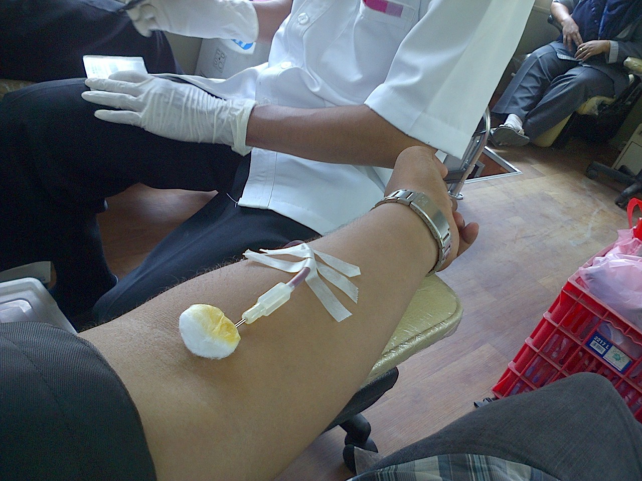
Thus, in order to analyze inflammasome activation in a more “in vivo”-like setting we optimized and validated a low-volume human whole blood assay (WBA) in this study using only 140 µl of whole blood per sample.
Therefore, we focused on the three well-characterized inflammasomes – NLRP3, AIM2, and NLRC4. Each of them was stimulated with a distinct agonist – in case of NLRP3, we performed a subsequent stimulation with ultra-pure lipopolysaccharide (up-LPS) and ATP. The NLRC4 inflammasome was stimulated with S.typhimurium, and, finally, the AIM2 inflammasome was stimulated via a transfection of double-stranded DNA, which, in our case, was poly (dA:dT).
We used IL-1β as a surrogate marker for inflammasome activity and measured IL-1β-levels in the supernatant of our whole blood samples after the end of stimulation by use of bead-based ELISA.
In case of each inflammasome, we were able to determine a decent amount of IL-1β after respective stimulation, concluding that inflammasome activity can be measured after stimulation in low-volume human whole blood samples.
Interestingly, in case of the NLRP3-specific stimulation with just up-LPS alone, we could already measure increased IL-1β-levels, which could be explained by a constitutively active caspase-1 due to an alternative NLRP3 pathway specific to only human monocytes.
In order to verify the accuracy of our chosen stimulation, we furthermore included various inhibitors into our assay that are described to be inflammasome-specific. High-extracellular potassium, as well as the NLRP3-specific inhibitor MCC950, were able to significantly diminish IL-1β-secretion in our assay and thereby underline the inflammasome-specificity of our chosen NLRP3-stimulation.
Approaching the AIM2 inflammasome, we dealt with several standard transfection protocols using different transfection agents as well as different durations of transfection. Concerning IL-1β, our best results could be obtained after a first stimulation (“priming”) with up-LPS similar to our NLRP3-stimulation and thereafter transfection using Lipofectamine for 6 hours. So, altogether, we designed an assay that lasts approximately 12 hours and leads to sufficient IL-1β-secretion after AIM2 stimulation.
Again, we also included the mentioned inhibitor. While extracellular potassium is able to completely inhibit IL-1β-secretion dose-dependently, in the case of MCC950, which is described to be exclusively NLRP3-specific, we only observed a partial inhibition. These findings led to the conclusion that our chosen AIM2 stimulation led to a simultaneous activation of NLRP3 and AIM2 inflammasome.
The third investigated Inflammasome was NLRC4. During our study, we observed a time-dependency in the concentration of secreted IL-1β after stimulation with S.typhimurium, where higher concentrations could be measured after longer stimulation.
The use of extracellular potassium led, similarly to our AIM2-data, to a dose-dependent inhibition of IL-1β-concentrations. However, the effects were stronger after shorter stimulation. Specific inhibition of the NLRP3-inflammasome using MCC950 revealed a strong inhibition of IL-1β secretion at early time points. For longer stimulations, the inhibitory effect of MCC950 on IL-1β secretion faded, thereby uncovering an increased NLRC4 dependency.
Whole blood assays can be confounded by several factors. First of all, the choice of anticoagulant plays an important role since the often-used heparin blocks transfection of DNA, thereby heavily impairing activation of the AIM2 inflammasome. This led to the use of hirudin for all experiments. Furthermore, a decent agitation during stimulation is necessary to prevent sedimentation of the blood cells, which inhibits NLRP3-activation. Especially considering our assay as a possible future diagnostic tool, we also checked the influence of storage time of drawn blood samples before the start of the experiment. Surprisingly, storage time at room temperature up to 24h led to robust IL-1β-secretion for all examined inflammasomes, opening the future possibility for the delivery of blood samples to diagnostic facilities.
Using the NLRP3 inflammasome specific inhibitor MCC950, we are able to specifically analyze NLRP3, AIM2, or NLRC4 inflammasome signaling. The optimized assay needs only low-volume whole blood samples, is independent regarding the hirudin concentration used and shows relatively robust IL-1β signals, even after prolonged storage of the whole blood samples. Therefore, the assay may be suitable for clinical research applications and even in pediatric patients. Given the fact that inflammasome-related signaling pathways are linked to a multitude of disease-causing pathologies, WBAs capable of deciphering specific inflammasome pathways could help to understand their molecular pathophysiology.
These findings are described in the article entitled An optimized whole blood assay measuring expression and activity of NLRP3, NLRC4 and AIM2 inflammasomes, recently published in the journal Clinical Immunology. This work was conducted by Lev Grinstein, Kristin Endter, Christian M. Hedrich, Sören Reinke, Hella Luksch, Felix Schulze, Angela Rösen-Wolff, and Stefan Winkler from Technische Universität Dresden, and Avril A.B. Robertson and Matthew A. Cooper from the University of Queensland.


