The midbrain, also called the mesencephalon, has multiple functions. These functions are the regulation of temperature, control of vision and hearing, motor control, controlling the sleep-wake cycle, and arousal. The brain operates with assistance from the cerebral cortex, the cerebellum, and the substantia nigra. The substantia nigra is part of the midbrain that is linked to the motor system located in the basal ganglia.
The midbrain is located above the hindbrain, the cerebral cortex, and situated near the center of the brain overall. The brain and spinal cord link together to enable the various functions of the midbrain. Voluntary movements are triggered by the rubrospinal tract, which runs from the cerebellum downwards to the spinal cord. The midbrain is comprised of many different parts. A closer look at the structure and function of the midbrain will help contextualize its role within the brain as a whole.
Making Sense Of The Midbrain’s Functions
Your midbrain is what allows you to sit up at your computer or to look at your phone and read this. Your midbrain is what lets you coordinate your actions and balance yourself, and it plays a major role in both auditory and visual reflexes. The midbrain is also responsible for the regulation of dopamine production. Dopamine is the “motivation” chemical of the body and it is responsible for our habits, behavior, attention, movement, and mood. The midbrain also helps control our sleep/wake cycle, regulating our alertness.
The midbrain must be able to do all these things for us to survive. Without the function of the midbrain, we wouldn’t be able to respond to threats or even move. For instance, if you accidentally touch your hand to a hot stove, your midbrain is what lets you jerk your hand back. The midbrain is what controls your motor movement and reflexes, letting you respond appropriately to situations like that.
Parts Of The Midbrain
The midbrain can be divided into four different parts or regions. These regions are the tegmentum, the tectum, the cerebral aqueduct, and the cerebral peduncles.

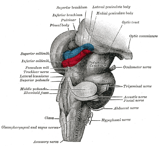
The tectum and its connections. Photo: By Henry Vandyke Carter – Henry Gray (1918) Anatomy of the Human Body (See “Book” section below)Bartleby.com: Gray’s Anatomy, Plate 719, Public Domain, https://commons.wikimedia.org/w/index.php?curid=526909
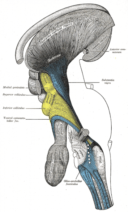
The tectum is the term given to the midbrain’s dorsal side, and it plays a role in reflex actions responding to auditory or visual stimuli. The tectum also gives inputs to the reticular spinal tract, which helps regulate our level of alertness. Four lobes or lumps found on the tectum are referred to as the corpora quadrigemina. The upper or superior to lobes are called the superior colliculi and they are responsible for the processing of visual information. They also help control certain eye movements and interact with fibers of the optic nerve. The superior colliculi are linked with the nerves located in the neck. Both of the superior colliculi are linked to a respective lateral geniculate nucleus. Near the superior colliculi are the inferior colliculi, which are responsible for the processing of auditory information and are found just above the trochlear nerve. Much like the superior colliculi, the inferior colliculi are connected to the corresponding medial geniculate nucleus, and they send information to them.
The cerebral aqueduct runs from the third ventricle to the fourth ventricle, as it is part of the ventricular system. The cerebral aqueduct is the ventricular system’s smallest ventricle, but it plays an important role in continuing the flow of the cerebrospinal fluid. Right cerebral aqueduct is surrounded by the periaqueductal gray and is found in between the tegmentum and the tectum. The periaqueductal grey is responsible for bonding, analgesia, and quiescence among other things.

The superior and inferior colliculi are in the yellow regions. Photo: By Henry Vandyke Carter – Henry Gray (1918) Anatomy of the Human Body (See “Book” section below)Bartleby.com: Gray’s Anatomy, Plate 685, Public Domain, https://commons.wikimedia.org/w/index.php?curid=541493
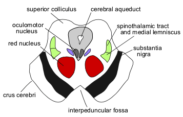
The cerebral aqueduct contains the nuclei of two pairs of cranial nerves, the oculomotor nuclei and the trochlear nuclei. The oculomotor nuclei are responsible for controlling most eye movements and including the movement of the eyelids. The oculomotor nuclei are found alongside the superior colliculus. In contrast, the trochlear nuclei are found at the level of the inferior colliculus and they help refine vision, focusing the eyes on proximal objects. The oculomotor nerve runs the ventral width of the tegmentum, emerging out of the nucleus. Similarly, the tectum is also near the point of emergence for the trochlear nerve. The trochlear nerve exits the brainstem dorsally, being the only cranial nerve to do so. The size of the pupil, as well as the shape of the lens, is controlled by a structure known as the Edinger-Westphal nucleus, found between the cerebral aqueduct in the oculomotor nucleus.
The tegmentum is substantially larger than the tectum, and it is located ventral to the cerebral aqueduct. The superior cerebellar peduncles enable the tegmentum to communicate with the cerebellum. These cerebellar peduncles are located at the level of the inferior colliculus, and in between the two peduncles is a structure called the median raphe nucleus, which assists other brain structures in consolidating memories. The main portion of the tegmentum is comprised of a complex system of neurons, forming a synaptic network that is involved with reflex actions and helping the body maintain homeostasis.
The tegmentum has many nerve tracts running through it as they move towards other parts of the brain, and it also has portions of the reticular formation within it. A small band of fibers referred to as the medial lemniscus are found running through an axial position near the inferior colliculus, while the spinothalamic tract is immediately dorsal to it. The red nuclei are structures which assist in motor coordination, and they are found at the level of the superior colliculus, and are red, round regions of the midbrain. The red nucleus has the nerves of spinal tract emerging from it, descending towards the spine’s cervical portion, where the red nuclei’s decisions about movement will be carried out. The ventral tegmental area is the area located between the two red nuclei on the ventral side. This structure plays a critical role in the neural reward system, being the region of the brain that produces the most dopamine. There are two parts of the forebrain connected with the ventral tegmental area, the hypothalamus, and the mamillary bodies.

Photo: Lipothymia, CC BY-SA 3.0, https://commons.wikimedia.org/w/index.php?curid=1228924
The basis pedunculi are found on either side of the brain’s midline, each comprising a lobe situated ventrally to the tegmentum. The Cris cerebri makes up the vast majority of the basis pedunculi. These are the main brain sections that drop down from the thalamus to the central nervous system. Some sections of the crus cerebri join the pons to the cortex, while the central and medial portions of the structure contain the corticospinal tracts. Be aware that the crus cerebral is also occasionally referred to as the cerbral peduncle, though this term actually encompasses all the fibers that transfer signals to the cerebrum.
The substantia nigra refers to a long dark band of the lobes that are connected to the tegmentum. Most of the basal ganglia is inside the forebrain, with the substantia nigra being the only portion that lies outside it. This structure is responsible for things like learning, processing reward stimuli, and motor planning. This region produces large amounts of melanin (responsible for its dark coloration), noradrenalin and dopamine.
Effects Of Midbrain Damage
A wide variety of problems can occur if the midbrain is damaged. Damage to structures in the midbrain can result in issues such as difficulty hearing, seeing, and recalling memories. Damage to certain areas of the midbrain has even been linked with Parkinsons’ disease.
The degeneration of the substantia nigra’s nerve cells can result in a reduction of dopamine levels and severely reduced dopamine levels are one of the suspected causes of Parkinson’s disease, which is a neurodegenerative disorder that causes problems with both coordination and motor control. Symptoms of the disease include slowed movement, tremors, trouble balancing and muscle stiffness.
Other Parts Of The Brain
The Forebrain
There are two other parts of the brain: the forebrian and the hindbrain. The forebrain is the largest part of the brain. The forebrain is made primarily of the cerebrum, which constitutes around two-thirds of the brian’s entire mass. The forebrain can be divided into two separate parts – the diencephalon and the telencephalon. The telencephalon is itself divided into four different lobes: the frontal lobe, the parietal lobe, the occipital lobe, and the temporal lobe. The frontal lobes are responsible for memory, thinking, planning, and voluntary muscle movement. The parietal lobes process sensory information, while the occipital lobes process visual information from the retina. The temporal lobes aid in the production of speech and the interpreting of language, as well as the formation of memories.
The diencephalon is a brain region which joins portions of the nervous system and endocrine system and relays sensory information between the two. The diencephalon is composed of the pineal gland, thalamus, and hypothalamus. The diencephalon plays a role in the regulation of motor functions, the endocrine system, and the autonomic functions.
The Hindbrain
The hindbrain is divided into the metencephalon and the myelencephalon. The metencephalon contains the cerebellum and the pons. The pons controls arousal, sleep, and various autonomic functions. The cerebellum helps control balance, equilibrium, fine, and coordination, and relays information between different muscles. The Myelencephalon contains the medulla oblongata, and it transmits sensory signals and motor signals between the higher brain regions and the spinal cord, which aids in the regulation of actions like sneezing, swallowing, breathing and heart rate.


