The function of Centrioles is to play a critical role in the orientation and attachment of microtubules to chromosomes during cell division. Centrioles function as the point of nucleation for the formation of the mitotic spindle during meiosis and mitosis.
That’s the short answer on the function of centrioles, but in order to fully understand the important role that centrioles have, it would be helpful to contextualize the role of the centrioles by taking a look at the processes of mitosis and meiosis.
The Structure Of Centrioles
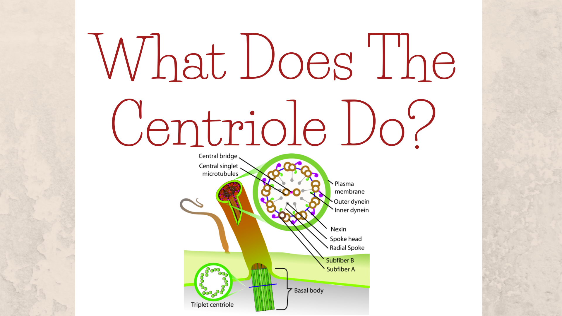
The centriole is made out of nine bundles of three microtubules arrayed in a circular fashion. The centriole has an appearance similar to a barrel, the daughter centriole, and the mother centriole are arrayed at a 90-degree angle to one another, giving the structure they form (the centrosome) a T-shape. The centrioles are surrounded by a thick matrix referred to as pericentriolar material. The diameter and length of the centrioles can change depending on the type of cell they exist in.
“The dream of every cell is to become two cells.” — Francois Jacob
Mitotic Cell Division

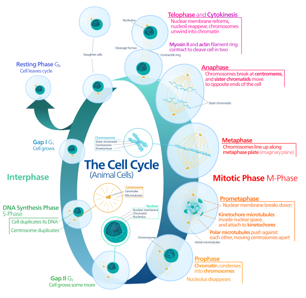
Photo: Kelvinsong via Wikimedia Commons, CC 1.0 – Public Domain
Mitosis is the process that divides one cell into two cells, allowing the cell to reproduce. Mitosis is divided into four or five different stages:
- Prophase
- Prometaphase (because prophase is so long it is sometimes divided into prophase and prometaphase)
- Metaphase
- Anaphase
- Telophase
By the time prophase starts the cell has already created a copy of its DNA, so within the nucleus of the cell are two structures called sister chromatids, copies of the chromosomes which are connected. A centriole binds with another centriole to create a structure called a centrosome, which is then itself copied. The centrosomes help organize long fibers called microtubules that will pull apart the chromosomes during the division of the cell.
The centrosomes move to opposite sides of the cell in prophase, and then the microtubules form a system of fibers called the mitotic spindle and join to the chromosomes and the centrosomes. The stretchy spindle widens as the centrosomes move to opposite poles.
“Life is the division of human cells, a process which begins at conception.” — Dick Gephardt
During the second part of prophase (prometaphase) the nuclear envelope that surrounds the nucleus breaks down and releases the chromosomes into the cytoplasm of the cell. The mitotic spindle that was created in between centrosomes no expands to capture the chromosomes. The microtubules bind to the chromosomes at a site known as the kinetochore, which is located at a region of the sister chromatids known as the centromere.
When metaphase beings the mitotic spindle that exists between centrosomes has bonded to the chromosomes and organized them, putting them into a linear horizontal arrangement at the center of the cell. The microtubules should anchor the chromosomes firmly to the centrosomes found on both sides of the cell. It’s important that chromosomes and centrosomes are attached in this way, otherwise, the chromosomes won’t properly divide during division. The cell double-checks to make sure this is the case. The cell goes through a process known as the spindle checkpoint, and if the sister chromatids are improperly aligned or attached cell division will be halted until the chromosomes can be properly aligned.
During anaphase of the cell, the chromatids come to the rest of the metaphase plate, not a true physical structure just a name for the region at the center of the cell. The two centrosomes (remember that each centrosome is comprised of two centrioles), begin to pull on the sister chromatids and draw them apart. The sister chromatids split apart into separate daughter chromosomes after they are pulled apart. The daughter chromosomes are then pulled by the mitotic spindle to opposite sides of the cell.
Note that the microtubules not involved in pulling the chromosomes apart begin to elongate during this part of the process, which has the effect of pushing the cell apart. This separates the poles from one another and elongates the whole cell to prepare for the division.
Telophase is the final portion of cell division, which occurs when the cell is almost finished dividing itself. The two halves of the cell that separate to become their own separate cells, and then begin re-building critical, regular cell structures like the nuclear membrane. The mitotic spindle will break down into its component parts and the chromosomes unwind. Once the process of cytokinesis is complete, two new cells have formed. Cytokinesis begins in either anaphase or telophase, depending on the type of cell undergoing division. The cells of animals divide by pinching the cytoplasm in the middle until the cells have divided, while the cells of plants simply create a new cell wall down the center of the cell.
The Process Of Meiosis
The primary difference between meiosis and mitosis is how the parent cells separate into daughter cells. While mitosis only has a single round of genetic separation and cellular division, meiosis has two different rounds of division and separation. The DNA found within sex cells, the cells which undergo meiosis, aren’t genetically identical to their parents, unlike the DNA found within cells that have undergone mitotic division.
“You are made of a hundred trillion cells. We are, each of us, a multitude.” — Carl Sagan
Sex cells go through the various phases as they divide, just like non-sex cells. The phases are the same as mitotic division: prophase, metaphase, anaphase, and telophase. Instead of these processes happening once, they happen twice, creating four daughter cells instead of only two daughter cells. Furthermore, each daughter cell has only one-half the number of chromosomes that the original cell does.
Between prophase and metaphase of Meiosis I, the homologous chromosome pairs randomly exchange their genetic information in a process dubbed “crossing over.” This crossing over process results is what results in the creation of non-identical chromatids. The cells are haploid cells, meaning that they only have one complete set of chromosomes, unlike diploid cells that have two sets of chromosomes. Meiosis II has the daughter cells divide again, which creates four haploid cells with non-duplicate chromosomes since the chromosome orient themselves for division randomly during this phase. These haploid cells end up becoming egg cells and sperm cells.
Throughout this process, centrioles play the exact same role they play in the mitotic division process.


