


The parts of the human heart can be broken down into four chambers, muscular walls, vessels, and a conductive system. The two upper chambers are called the atria, with lower parts called ventricles. These all work together to make up the vital function of your heart.
Everybody knows that the human heart is the essential organ in our bodies. But few people know all its parts and their essential functions.
Broadly speaking, the human heart has one function: to pump blood through the circulatory system all throughout the body, thus supplying both nutrients and oxygen to the body’s tissues while also removing all the wastes, including carbon dioxide.
The Anatomy of the Human Heart
Here are some important facts about the anatomy of the human heart:
- The human heart can weight between 200 to 425 grams (or 7 and 15 ounces). On average it beats about 100,000 times a day and pumps 7,571 liters (2,000 gallons) of blood.
The medical literature tells us that the most effective ways to reduce the risk of heart disease, cancer, stroke, diabetes, Alzheimer’s, and many more problems are through healthy diet and exercise. Our bodies have evolved to move, yet we now use the energy in oil instead of muscles to do our work – David Suzuki
- Most people are not aware of the exact location of the heart. It is between the lungs, approximately in the middle of the chest, right behind the sternum (breastbone) but slightly to the left.
- The human heart beats (contracts) each time it received an electrical impulse from the heart muscle, known as the myocardium.
- The human heart together with the circulatory system make up the cardiovascular system. The blood that your heart pumps gets to every single cell in your body and delivers nutrients and oxygen to each of them while also removing all the waste products including carbon dioxide.
What Are the Parts of the Human Heart?
Here is the full list of all the parts of the human heart and their essential functions:
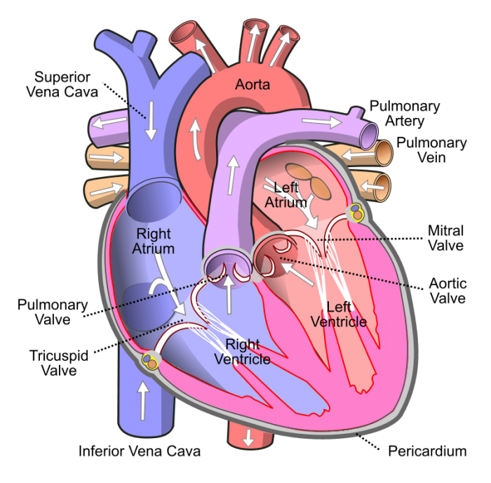
Photo: By Wapcaplet – Own work, CC BY-SA 3.0, https://commons.wikimedia.org/w/index.php?curid=830253
The Aorta: this is the largest artery in the human body. Arteries are tubular branching elastic-walled muscle vessels that carry blood all the way from the heart through the body. The main function of the aorta is to take oxygenated blood all the way from the left ventricle to the rest of the body.
The Pulmonary Artery: this artery is responsible for carrying deoxygenated blood all the way from the right ventricle to the lungs.
Oxygenated blood: the word “oxygenated” simply means that it carries oxygen. So, oxygenated blood is the blood that carries oxygen.
Deoxygenated blood: the word “deoxygenated” simply means that it carries very little or no oxygen. So, deoxygenated blood is the blood that carries either very little or no oxygen.
The Right Atrium: this part of the human body is tasked with receiving deoxygenated blood from the rest of the body.
The Pulmonary Vein: Veins are the tubular branching vessels that carry blood all the way from the capillaries and take it to the heard. The function of the pulmonary vein is to take oxygenated blood all the way from the lungs to the left atrium.
The Left Ventricle: A ventricle is any of the cavities of a bodily part or organ. This ventricle, in particular, is the chamber of the heart that gets blood from a corresponding atrium. The left verticle is from where the blood is forced into different arteries. The essential function that the left ventricle carries out is pumping oxygenated blood into the aorta.
The Right Ventricle: The right ventricle is tasked with pumping deoxygenated blood into the pulmonary artery.
The Coronary Vessels: these vessels supply the myocardium (the heart muscle) with the necessary supply of blood. There is a main left coronary that goes into the circumflex artery, supplying blood to the left atrium. There is a right coronary vessel that goes into the right marginal artery, supplying blood to the right atrium and the right ventricle.
The Arteries: the arteries are tasked with carrying blood away from the heart. These blood vessels are muscular tubes. The aorta is the largest artery. Each and every artery is lined with three layers of sooth tissue. The three layers are the intima, which is the inner layer whose tissue is called endothelium; then, there the media, which is a muscle layer whose role is to allow the human heart to deal with high pressures; finally, there is the adventitia, which connects the arteries to tissue.
The Veins: the veins are tasked with carrying blood toward the heart.
The Bicuspid Valve: Valves are bodily structures (such as the mitral valve) that either shut down temporarily an orifice or passage or that permits fluid to move, but always in only one direction. The Bicuspid Valve is the valve located between the left ventricle and the left atrium. Some people are born with this kind of aortic valve that is located between the aorta and the left ventricle and has two cusps instead of the usual three. People who are born with a bicuspid valve instead of a tricuspid valve may be affected by it, particularly, once they become adults. This valve often causes what is known as aortic valve stenosis, which is the narrowing of the aortic valve. People with a bicuspid valve sometimes suffer from an enlarged aorta and this can increase the risk of having an aortic dissection.
The Tricuspid Valve: this is the vale that is located between the right ventricle and the right atrium. Most people are born with a tricuspid valve, which is the valve with three cusps located between the aorta and the left ventricle.
The Vena Cava: this is the largest vein in the human body (vena is Latin for vein). Its essential function is to carry blood from all around the body all the way to the heart. There is a superior vena cava and an inferior vena cava. The superior vena cava is tasked with carrying blood to the upper body: neck, head, and both upper limbs back to the heart. What the inferior vena cava does is carrying blood back from the lower parts of the body back to the heart.
Related Posts
 Important Strategies To Help Healthcare Providers Support Patients With Diabetes
 Studying The Link Between Increased BMI And Late-Onset Preeclampsia In Pregnant Women
 A Bacterial Cell Imaging Method Using CRISPR And Microfluidics
 B. infantis Reduces Key Markers Of Intestinal Inflammation In Infants
 Epigenetic Changes In Multiple Sclerosis – Studied In Twins
 Did You Know Math Can Help Us Learn How Diseases Work?