


In oncology, the development of new efficient treatments is still a challenge, even today. Indeed, besides the complexity to discover good anticancer candidates, the process is still too long and expensive, requiring an average of ten years of work and several million dollars of investment.
Before beginning drug tests on patients included in clinical trials, preclinical assays are needed to assess drug efficiency and toxicity. In the field of oncology, preclinical tests are usually realized on cancer cells (ex vivo model) or on animals (in vivo model). The ex vivo model offers the opportunity to screen a large panel of drug candidates but has the major disadvantage of not being representative of tumor complexity to include the pathophysiology, histology, and genomic mutations of tumors. Animal models are more representative of the tissue environment. However, drug tests frequently fail because of physiological differences with humans.
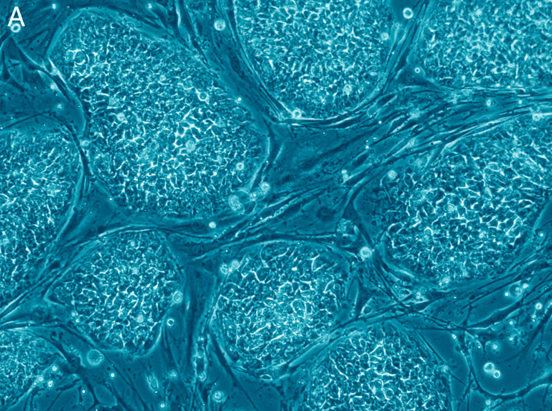
To address these limits, a new technology called organoïds has emerged as a promising model for preclinical drug tests. Organoïds are described as three-dimensional (3D) structures derived from stem cells, which, when they are grown in 3D conditions, can self-organize into architectures that mimic those of a miniaturized organ. Therefore, organoïds are more representative of the physiological organogenesis.
Another advantage of this model is that organoïds can be generated from stem cells isolated from human tissues. Stem cells are immature and unspecialized cells characterized by two properties which distinguish them from other cell types: the ability of self-renewing, producing the same cells and, under some conditions, the capacity to differentiate, producing mature and specialized cells (i.e blood cells, cardiac cells, muscle cells…).
Organoïds can be derived from 3 types of cell lines: (i) physiological stem cells, including embryonic stem cells, neonatal stem cells, and adult stem cells; (ii) induced stem cells, including the induced pluripotent stem cells (IPS), and (iii) cancer stem cells. For drug screening in oncology, organoïds can be derived from cells isolated from patient cancer biopsies. Nowadays, researchers have generated several organotypic structures including kidney, gut, brain, liver, pancreas, stomach, retinal tissue, prostate, and fallopian tubes. Therefore, tumors can be reproduced, giving a disease-specific model for drug tests. In addition, organoïds appear as a promising model to develop a personalized medicine. Indeed, organoïds can be specifically generated from one patient, then anticancer drugs or anticancer candidates can be tested on the generated organoïds offering the possibility of a patient-specific therapy.
Despite the many advantages of the organoïd model, this technology is still not perfect and needs to be improved. The production time remains too long and the cost too high. The automation of organoïd generation could answer this problem.
These findings are described in the article entitled Human-cell-derived organoids as a new ex vivo model for drug assays in oncology, recently published in the journal Drug Discovery Today. This work was conducted by Miryam Mebarki, Annelise Bennaceur, and Laurence Bonhomme-Faivre from the APHP-Hopital Paul Brousse.
Related Posts
 Important Strategies To Help Healthcare Providers Support Patients With Diabetes
 Studying The Link Between Increased BMI And Late-Onset Preeclampsia In Pregnant Women
 A Bacterial Cell Imaging Method Using CRISPR And Microfluidics
 B. infantis Reduces Key Markers Of Intestinal Inflammation In Infants
 Epigenetic Changes In Multiple Sclerosis – Studied In Twins
 Did You Know Math Can Help Us Learn How Diseases Work?