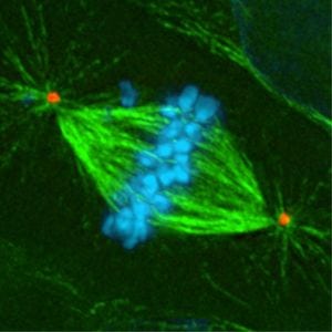
Microtubules play a crucial role in segregating chromosomes from one cell to another during cell mitosis and division. They function as a skeleton to give cells their shape and enable many biological processes such as cell to cell communication and protein transport within cells.
Microtubules were first observed in the sea-urchin egg in 1928 (Runnbtrc-M, 1928). At that time scientists were using the term “mitotic figures” to describe microtubules because they were observed during metaphase of mitosis. The same “mitotic figures” were found in plants (1934), in the cells of a chick (1948), in worms (1950), and many more. Using early imaging techniques, scientists observed these structures as being most pronounced during metaphase but then gradually disappearing during later stages of cell division. Scientists did not know what to make of the formation and breakdown of these figures and thought that the intrinsic forces generated in dividing cells caused chromosomes to move to opposite sides of daughter cells.
Using early imaging techniques, scientists observed that these structures were clearly pronounced during metaphase, but then appeared to gradually disappear during later stages of cell division. Scientists did not know what to make of the formation (polymerization) and breakdown (depolymerization) of these figures and thought that an unknown, but inherent, natural force caused chromosomes to move to opposite sides of a dividing cell to be distributed to the daughter cells. The advancement of imaging techniques by 1963 led to the observation of mitotic figures being dispersed throughout a cell’s cytoplasm, rather than just being present during metaphase. Due to their rod-shaped structure, they were then given the name “microtubules.” Four years later, tubulins, the building blocks of microtubules, were identified.
From the knowledge gained through pioneering studies in both simple and complex model organisms such as these, we now know that microtubules interact with chromosomes during the entire cell cycle, not just during cell division. Newly duplicated chromosomes must be divided equally between daughter cells during cell division, but there is no inherent “force” generated in dividing cells. Microtubule filaments composed of tubulins are now known to play an active role in chromosome separation. These microtubules function as the “skeleton” of cells which give cells their shape and maintain their integrity.
Microtubules also enable many biological processes, such as protein transport within cells and cell to cell communication. These basic cellular functions are essential for survival. Unfortunately, cells do not function perfectly all the time and can make mistakes, some of which create diseases and hijack cellular components. For instance, when a cell does not follow the built-in program for cell division, the cell may lose control of the cell division process. This loss of control can result in premature cell death. Alternately, too-frequent cell division can cause cancer in the host organism.
Based on this research, you might understand how important microtubules are for normal biological processes. But a pertinent question remains: how do tubulins organize themselves into microtubule filaments? At that time, there were only two known types of tubulins that make up microtubules: alpha and beta tubulin, and alpha and beta tubulins are just the building blocks that create microtubule filaments. Model organisms were once again critical in answering the question of how tubulins are organized into microtubule filaments in 1989 in the lab of Dr. Berl Oakley.
Dr. Oakley was interested in identifying proteins which associate with microtubules in the cytoplasm, particularly during the breakdown of microtubule filaments. Dr. Oakley and his team decided to use genetic screening, a classic genetic approach where random mutations in the genome of an organism are created using mutagens. The physical characteristics, or phenotypes, of the mutants are recorded, and a gene of interest is then isolated based on a particular phenotype. By using genetic screening on the common fungal model species Aspergillus nidulans, Dr. Oakley and his team targeted a gene that fit their screening criteria: one that seemed to interact with beta-tubulin. As an unknown tubulin gene, this gene had never been studied, so Dr. Oakley’s team set out to isolate and understand why this particular gene was important for microtubules. They were surprised to discover that this gene, which they named Gamma tubulin, was the foundation of microtubule formation. Specifically, gamma tubulin is the backbone where all other tubulins connect to form microtubule filaments.
Knowing and understanding the molecular components and cellular mechanisms of how microtubules are organized and the role they play in cell division led to scientists developing anticancer drugs that block microtubule formation in cells with uncontrolled cell division, once again demonstrating the power of model organisms in advancing science and medicine. An initially unexplained observation of the presence of mitotic figures in the egg cells of sea-urchins was clarified through years of work in traditional model organisms, such as plants and worms, but also in less traditional model organisms, such as chicks and sea-urchins, and led to the significant discovery and characterization of tubulins and microtubule filament formation.
I would like to thank Sumeet Nayak from the University of Massachusetts Medical School and the Communication and Outreach Subcommittee of the Genetics Society of America for his contribution and input.
This article is based on research conducted by Haifa Alhadyian, an Early Career Scientist from the University of Kansas and a Communication and Outreach Leader at the Genetics Society of America. For more information about the journey behind the discovery of microtubules, please visit Microtubules: 50 years on from the discovery of tubulin.


