


The cornea is the first part of the eye that the light enters to allow us to see. It also acts as a barrier against the external environment, so it is very susceptible to wounding and injury. Given that improper wound healing in the cornea is the fourth-highest cause of preventable blindness according to the World Health Organization, wound healing in the cornea is an important subject of study. Furthermore, common diseases like type-2 diabetes can hinder this process, and it has been an increasing healthcare concern in the U.S. Our goal was to study the mechanism of how the cornea signals to the epithelium for wound healing.
In our past studies, we reported that when the cornea is wounded through scratch-wounding, ATP is released from the injured cells as a signaling molecule. This ATP stimulates two cell receptors, P2X7 and P2Y2, which generate Ca2+ signaling. This pathway was found to be important in mediating the wound healing process. The wound healing process coordinates all the epithelial cells in the cornea to move as a unit, suggesting that there is communication between cells that occur during the process. However, the behavior of this Ca2+ in the cornea cells has not been fully characterized.
The goal of our study was to develop a computer program system to identify and characterize the Ca2+ cell-cell communication in the cornea. We used MATLAB to do this, and we were able to characterize the different Ca2+ signaling patterns in P2X7 and P2Y2. The presence of Ca2+ signaling in adjacent cells were also seen to be directly associated with changes in cell shape and movement. Overall, the cells at the front of the wound acted like “leader cells” that initiated the cell movement needed for wound healing. We were able to show this in both corneal cell lines and in mouse cornea tissue. Together, our results demonstrate that these Ca2+ signals are a vital component in regulating the long-term response to injury in the cornea.
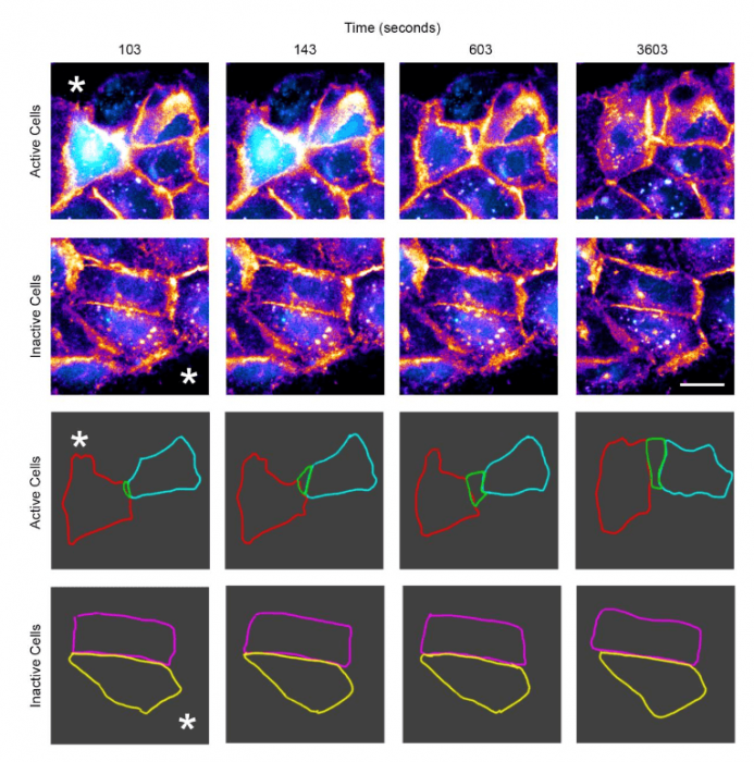
Figure republished with permission from PLOS from https://doi.org/10.1371/journal.pone.0213422
These series of images show how Ca2+ signaling between cells correlate with changes in cell shape and movement. The corneal epithelial cells were preincubated with a membrane dye (in orange) and a Ca2+ indicator dye (cyan) to visualize the cells. The injury was made by scratch-wounding (indicated in asterisks) and cells that exhibited Ca2+ signals (active cells, first row) showed drastic changes in cell motility and cell shape compared to cells that did not have those mobilizations (inactive cells, second row). The same cells were tracked by outlining them over time (third and fourth row) to observe the changes.
These findings are described in the article entitled Sustained Ca2+ mobilizations: A quantitative approach to predict their importance in cell-cell communication and wound healing, recently published in the journal PLOS One.
Related Posts
 Important Strategies To Help Healthcare Providers Support Patients With Diabetes
 Studying The Link Between Increased BMI And Late-Onset Preeclampsia In Pregnant Women
 A Bacterial Cell Imaging Method Using CRISPR And Microfluidics
 B. infantis Reduces Key Markers Of Intestinal Inflammation In Infants
 Epigenetic Changes In Multiple Sclerosis – Studied In Twins
 Did You Know Math Can Help Us Learn How Diseases Work?