


The Golgi body, also sometimes referred to as the Golgi apparatus or Golgi complex, is an intracellular organelle that is responsible for the packaging and transport of protein products. Proteins that are manufactured in the endoplasmic reticulum (ER) are sent to the Golgi body, where they are then modified and sent off for secretion via exocytosis. In many ways, the functioning of the Golgi apparatus can be likened to that of a post office.
Just as a post office organizes, labels, and sends off packages, the Golgi apparatus functions to organize, label, and send off proteins to the proper intra- or extracellular locations.
“The body is a cell state in which every cell is a citizen. Disease is merely the conflict of the citizens of the state brought about by the action of external forces.” — Rudolf Virchow
Due to its size and unique shape, the Golgi complex was among the first cellular organelles to be properly identified and observed. Perhaps ironically, it was also one of the last organelles to have its existence definitively confirmed. It was first discovered by Italian physician and neurologist Camillo Golgi in 1898 when Golgi observed the organelle via his own idiosyncratic method for staining neural cells. At that time, Golgi named the newly discovered organelle apparato reticolare interno (“internal reticular apparatus”). For almost 50 years afterward, fellow physicians thought the organelle did not actually exist; they considered its “discovery” to simply be an optical illusion resulting from the particular staining techniques Golgi used. It was not until the 1950s that microbiologists confirmed the reality of the organelle, and gave it its present name of the Golgi body in honor of its discoverer.
Structure Of The Golgi Body
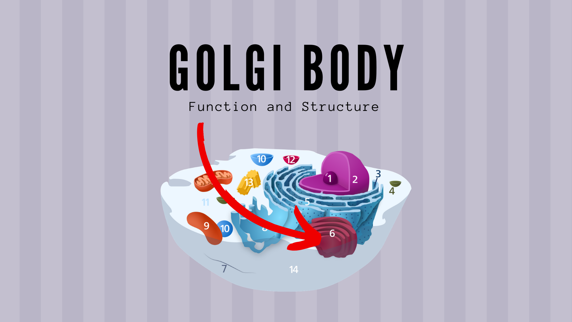
In eukaryotic cells, the Golgi apparatus is one of the cell’s larger organelles, coming in at about 200 nm in length. The Golgi body can also modify its size in response to the performance needs of the cell. The organelle is composed of a series of flattened membrane-enclosed discs called cisternae. Typical mammalian cells contain anywhere from 40-100 of these cisternae.
“A cell of a higher organism contains a thousand different substances, arranged in a complex system.” — Herbert Spencer Jennings
This collection of cisternae are further divided into two functional pathways, the cis-Golgi network (CGN) and the trans-Golgi network (TGN). These systems correspond to the “entrance” and “exit” of the Golgi body, respectively. The CGN composes the first system of the Golgi body and is responsible for the intake and initial processing of proteins. The TGN is located at the end of the packaging train and is responsible for packaging the proteins into vesicles to send off to other regions of the cell. These functional pathways are general features of the Golgi body but there are structural and organizational differences in the Golgi body between eukaryotes. A general rule though is that plant cells tend to have smaller but more numerous cisternae while animal cells have fewer but larger cisternae. In addition, the more secretions a cell produces, generally the larger its Golgi body is.
Function Of The Golgi Body
The Golgi body is primarily responsible for the individual packaging and transport of proteins that are synthesized by the endoplasmic reticulum. Once a protein is created in the endoplasmic reticulum, it is sealed in a vesicle and sent to the Golgi body. The vesicle bonds to the cis-face of the organelle, and transfers the contained protein over via carrier molecules. Once inside the Golgi body, the protein goes through “processing” where it is modified and marked for transportation. At the trans-face of the organelle, the processed protein is repackaged into a vesicle and sent off in its intended direction.
The majority of protein “processing” in the Golgi body is in the form of post-translation modification — the addition of extra-functional or chemical groups onto a protein that was synthesized via mRNA transcription. Essentially, the Golgi apparatus adds extra chemical groups to different parts of the protein. These chemical groups serve as markers that let the rest of the organelle know where to send the proteins. For example, lysosomal proteins are modified by the addition of a phosphate group to the protein’s sugar groups. Other common mechanisms of post-translational modification include the addition of carbohydrate and sulfate groups.
Once the proteins have been appropriately modified, they are packaged in vesicles and sent out to different parts of the cell. Depending on the chemical sequences added to the protein in the modification process, the TGN will package the protein in one of three vesicles, each corresponding to a different location. There is still a live scientific debate about the exact nature of the physiological mechanisms that underly vesicular trafficking and transport of the Golgi body.
The function of the Golgi body can perhaps best be understood by explaining what would happen without the Golgi body. If cells did not have a Golgi apparatus, they would be unable to differentiate which proteins are which and which proteins belong where. Imagine if the local post office suddenly disappeared. No one would be able to send packages or mail and there would be no mechanisms to mark and distribute packages or mail to the correct recipients.
Due to the wide variety of proteins that the Golgi body handles, pathologies of the Golgi apparatus can have a number of symptoms depending on which protein sequences are involved. For example, Pelizaeus-Merzbacher disease is a condition where mutations in the genes responsible for PLP1 production cause the protein to fold improperly and become stuck in the ER-Golgi apparatus pathway. Associated symptoms include ataxia, delayed motor development, and muscle atrophy. Alternatively, Parkinson’s disease has been associated with a build-up in a protein α-synuclein and a fragmenting of the Golgi apparatus. It is thought that the fragmenting of the Golgi apparatus prevents the processing of α-synuclein, the build-up of which can contribute to the neurodegenerative symptoms that characterize Parkinson’s disease.
“I don’t have any choice whether or not I have Parkinson’s, but surrounding that non-choice is a million other choices that I can make.” — Michael J. Fox
So in summation, the Golgi body is important for cell functioning as it serves to properly package, mark, and send of proteins for both intra- and extracellular use. Without a Golgi complex, the various proteins manufactured by the cell would float around aimlessly, completely powerless to perform their function.
Related Posts
 Important Strategies To Help Healthcare Providers Support Patients With Diabetes
 Studying The Link Between Increased BMI And Late-Onset Preeclampsia In Pregnant Women
 A Bacterial Cell Imaging Method Using CRISPR And Microfluidics
 B. infantis Reduces Key Markers Of Intestinal Inflammation In Infants
 Epigenetic Changes In Multiple Sclerosis – Studied In Twins
 Did You Know Math Can Help Us Learn How Diseases Work?