The parts of a human leg consist of bones, muscle, tendons, and ligament all working together so that your leg can perform complex movement. The leg parts can anatomically be divided into the crus, which is anything below the knee and the thigh which is anything above the knee. The crus of the leg can be then divided into the sural, or the calf, the peroneal or the side of the human leg and the shin or front of your leg.
People use their legs to get around all day, but most of them don’t stop to think about how their legs work and how the parts of their legs collaborate to enable motion. What are the various parts of the human leg, and what are the different roles these parts fulfill?
The meaning of leg differs depending on who you talk to. Colloquially, the leg refers to the entire lower limb of the body. This could include the entire limb, up to the thighs or even hips. Yet in anatomical texts, the leg usually only refers to the lower portion of the limb extending down from the knee to the ankle.
The Skeleton of the Leg
The skeletal system of the legs is what supports the muscles found in the legs, acting as the physical base for your entire leg. The major bones found in the leg are the femur, the tibia, the fibula, and the patella. The femur is the longest bone in the limbs of the body. It connects the hip bone to the knee. The femur is dense and compact since it must support the movement of the body. The patella, or kneecap, fits over the ligaments that join together the femur with the tibia and fibula. The patella protects the muscles and ligaments inside the joint of the knee.
In the bones of the lower leg, the tibia and fibula run from the knee to the ankle. There is a layer of cartilage called the meniscus between the tibia and the femur. The tibia is responsible for bearing most of the weight of the body, and the role of the fibula is to help balance the lower leg by supporting the lower leg muscles. The ankle joint is formed by the conjunction of the tibia with the tarsal bones found in the foot.
“How long should a man’s legs be? Long enough to touch the ground.” — J. D. Salinger
The Muscles of the Leg

Photo: Public Domain
The muscles in the human leg are what allow the leg to move. They sit on top of the bones and receive instructions to either relax or contract from the brain. They are supported by veins and arteries which deliver blood to them.
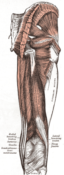
The muscles of the leg can be divided into two different groups: superficial muscles and deep muscles. The superficial muscles are the outside muscle layer while the deep muscles are the inner muscle layer.
In the upper leg above the knee, the major superficial muscles include the biceps femoris muscles, the semitendinosus muscles, and the semimembranosus muscle. The deep muscles of the thigh include the vastus lateralis muscle, the vastus intermedius muscle, and the vastus medialis muscle.

Photo: Public Domain
The superficial muscles include the gastrocnemius, the plantaris and the soleus muscles. All of these muscles run into the calcaneus of the foot. The gastrocnemius is a flexor for the knee, and so is the plantaris. The soleus muscle flexes the foot via the ankle joint. The deep muscles of the lower leg include popliteus, the tibialis posterior, the flexor digitorum longus, and the flexor hallucis longus. The popliteus muscle rotates the femur around the tibia, while the tibialis posterior flexes the foot itself, pulling up on the arch of the foot. The digitorum longus runs to the toes and flexes the smaller four toes while the hallucis longus flexes the big toe.
Skeletal muscles are either flexor or extensors. Flexors work by bending a joint, relaxing the muscle and pulling it back. Extensors work to extend a joint, contracting the muscle and allowing the body to extend itself.
Veins and Arteries
The muscles and other tissue in the legs are served by arteries and veins. The veins and arteries carry blood to the tissue in the legs, providing cells there with oxygen they need to carry out their functions. There are two main arteries that run from the area near the kidneys and down the length of the leg. These arteries are called the common iliac arteries, and there is both an internal and external iliac artery. The major artery in the thigh, the femoral artery, turns into a series of smaller arteries and veins as it continues down into the leg. Other important arteries in the legs include the popliteal artery, the posterior tibial artery, the anterior tibial artery, and the plantar arteries.
Much like the muscle groups, there are both deep veins and superficial veins in the legs. The deep veins are connected to the superficial veins via perforator veins. The deep veins in the body handle approximately 85% of returning blood flow, while superficial veins handle the other 15%. Superficial veins include the greater saphenous vein and the small saphenous vein. Deep veins in the leg include the femoral vein, the popliteal vein, the fibular vein, and the anterior and posterior tibial veins.
Nerve Pathways In The Legs

Photo: Public Domain
The nerve impulses carried by the pathways in the legs are what allow a person’s legs to move. The signals originate in the brain where they are carried across the synapses of neurons. They are then carried by the spinal cord down to the nerve clusters and pathways of the legs, which then stimulate the muscles and produce movement. There are a number of different stretch receptors in the legs, disperse throughout the muscles, tendons, ligaments, and joints.
The nerves that are found in the foot and leg come from the spinal cord in the lower back. As the nerves move towards the thighs and upper leg they branch off into two main networks called the sacral plexus and lumbar plexus. The lumbar plexus itself further divides into other nerves, like the femoral, obturator, and lateral femoral nerves. These nerves run into the skin and muscles of the leg, providing information on a number of sensations like pressure, pain, and temperature. Parts of the femoral nerve branch run all the way to the foot.
The sciatic nerve is one of the longest nerves in the body, and it runs down from the sacral plexus in the butt and thighs to deliver electrical impulses to the lower legs and feet. It divides into various nerve groups like the tibial and fibular nerves along the way.
The anatomy of the human leg is complex and multi-layered, requiring all of its parts working together to enable motion.


