


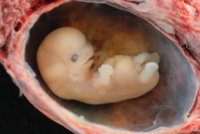
Embryonic development is a fascinating process where a fertilized oocyte develops into a multicellular, adult organism. For more than 70 years, it was thought that many morphological changes we observe during embryogenic development were driven by apoptosis, a form of cell death that sculpts our tissues and removes superfluous structures that are no longer required. Perhaps the best-known examples include the shedding of a tadpole’s tail during metamorphosis and the separation of our fingers and toes that initially developed as paddle-like structures with webs between the digits. Apoptosis was also postulated to be fundamental for shaping the embryo through invagination, evagination, fusion, and cavity formation.
To elaborate, apoptosis is a normal physiological process whereby a cell is programmed to undergo suicide. It is tightly regulated by the BCL-2 family of proteins, which consists of members that function to either promote cell death or, conversely, safeguard cell survival. The intricate balance between these proteins with opposing functions determines whether a cell will continue to live or die. Apart from morphogenesis, apoptosis is also responsible for eliminating diseased cells and maintaining homeostasis by removing old cells to counter-balance the production of new ones.
Two key players that are essential for driving apoptosis are BAX and BAK, functionally redundant pro-death proteins that puncture holes in the outer mitochondrial membrane, thereby committing cells irreversibly to death. This step, known as mitochondrial outer membrane permeabilization (MOMP), is considered the point of no return, because it results in the release of molecules that activate enzymes, which in turn cleave cellular DNA and proteins, leading to cell demolition. Therefore, it came as a great surprise when mice engineered to lack both BAX and BAK (also referred to as Bax-/-Bak-/- double knockout mice) were able to survive to birth, and a small number of these animals even developed to adulthood with relatively minor abnormalities, such as interdigital webbing. This led to the hypothesis that an additional protein may also harbor functions that overlap with those of BAX and BAK, which therefore allowed apoptosis to proceed in the cells of these Bax-/-Bak-/- double knockout mice.
A prime candidate was BOK, a poorly-studied member of the BCL-2 family that has a very similar amino acid sequence to both BAX and BAK. BOK was postulated to exert a pro-apoptotic role; however, its physiological function remained a highly debated topic until recently. In Embryogenesis and adult life in the absence of intrinsic apoptosis effectors BAX, BAK, and BOK, Ke and colleagues removed all three genes encoding Bok, Bax, and Bak to create Bax-/-Bak-/-Bok-/- these mice. The rationale behind this experiment was that if BOK was indeed the protein responsible for driving apoptosis in Bax-/-Bak-/- double knockout mice, the combined loss of BOK, BAX, and BAK would result in a more severe block in apoptosis, and predictably more severe developmental defects during embryogenesis.
The study revealed several unexpected outcomes. Firstly, while some Bax-/-Bak-/-Bok-/- these embryos failed to develop normally and displayed more severe defects than those seen in Bax-/-Bak-/- mice as anticipated, it was surprising to see that a large proportion of the these animals successfully completed the entire gestation period and were born. However, most of these pups died soon after birth due to a range of severe anomalies, in particular, cleft face, cleft palate, exencephaly, and heart vessel defects. Remarkably, however, many organs including the liver, stomach, and lungs formed normally in the Bax-/-Bak-/-Bok-/- these mice. Although some Bax-/-Bak-/- double knockout embryos and newborn pups also displayed similar defects, it was clear that these anomalies occurred significantly less frequently, and were also a milder form of the condition, which thus permitted a higher rate of survival.
To confirm that no apoptosis was occurring in cells from Bax-/-Bak-/-Bok-/- triple knockout embryos, the team also enumerated apoptotic cells in these embryos and confirmed that there were no detectable dying cells when BOK, BAK, and BAX were removed. Perhaps the most surprising discovery was that approximately 1% of the Bax-/-Bak-/-Bok-/- these mice had no obvious abnormalities, apart from webbed paws, and even survived to adulthood. These findings demonstrated that many events postulated to be apoptosis-dependent during embryogenesis still occurred in its absence, implying that apoptosis plays a restricted and supportive role during development. The manifestation of more frequent and more severe defects when all three genes (Bok, Bax, and Bak) were removed compared to the deletion of two genes (Bax and Bak) suggests that BOK has some functional overlap with BAX and BAK. Accordingly, by using X-ray crystallography the team confirmed that BOK is structurally similar to BAK and BAX, which provided a further piece of evidence that BOK indeed functions as a pro-apoptotic protein.
To conclude, Ke et al. showed that although apoptosis is critical during development, the failure of which can lead to severe defects and mortality, it does not play as widespread a role as previously thought. This study redefined the tissues that genuinely require apoptosis, including the brain, retina, digits, and palate, and provided an insight to how birth defects such as cleft palate, spina bifida, and exencephaly may arise.
These findings are described in the article entitled Embryogenesis and Adult Life in the Absence of Intrinsic Apoptosis Effectors BAX, BAK, and BOK, recently published in the journal Cell. This work was conducted by Francine F.S. Ke, Hannah K. Vanyai, Angus D. Cowan, Alex R.D. Delbridge, Stephanie Grabow, Peter E. Czabotar, Anne K. Voss, and Andreas Strasser from The Walter and Eliza Hall Institute of Medical Research and the University of Melbourne, and Lachlan Whitehead from The Walter and Eliza Hall Institute of Medical Research.
Related Posts
 Important Strategies To Help Healthcare Providers Support Patients With Diabetes
 Studying The Link Between Increased BMI And Late-Onset Preeclampsia In Pregnant Women
 A Bacterial Cell Imaging Method Using CRISPR And Microfluidics
 B. infantis Reduces Key Markers Of Intestinal Inflammation In Infants
 Epigenetic Changes In Multiple Sclerosis – Studied In Twins
 Did You Know Math Can Help Us Learn How Diseases Work?