Understanding the frequency components of brain signals and their functional relevance has been a central focus in neuroscience research. Therefore, since the inception of resting-state functional magnetic resonance imaging (fMRI), deciphering the seemingly noisy spontaneous fMRI oscillations has been critical. However, because of the nature of the hemodynamic response and low sampling rate of resting-state fMRI signals compared to electroencephalogram (EEG) or magnetic electroencephalogram, the exact composition of resting-state fMRI oscillations and their functional relevance remain controversial.
Yang and colleagues (2018) at the Center of Dynamical Biomarker at Beth Israel Deaconess Medical Center/Harvard Medical School, used the Hilbert-Huang Transform (HHT), a well-cited signal decomposition method to identify intrinsic component of resting-state fMRI signal in a large normal-aging cohort comprised of more than 400 subjects aged 21 to 89 years.
HHT is distinct to Fourier-based methods (or commonly used Fast Fourier Transform, FFT), HHT holds no priori assumption for underlying structures of the signal and is, therefore, an effective method for analyzing nonlinear and non-stationary signal consisting of multiple periodic components. This is an important advantage when analyzing brain signal because brain activity is known to have complex oscillations with their frequencies and amplitude change over time.

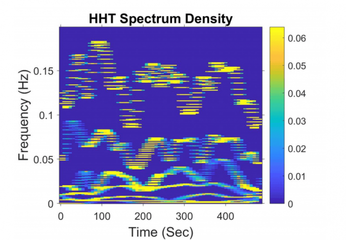
Fig 1. An example of Hilbert-Huang Transform of a resting-state fMRI signal (time points = 200 and sampling rate = 0.4 Hz). The time-frequency spectra show that resting-state fMRI signal consists of several intrinsic components with modulated frequencies over time. Image republished with permission from Elsevier from https://doi.org/10.1016/j.neurobiolaging.2018.06.007
The study revealed that resting-state fMRI power spectra consist of three bands: high (0.087–0.2 Hz), low (0.045–0.087 Hz), and very low (≤ 0.045 Hz) frequency bands. Yang and colleagues further tested the correlation of special power in these bands with cognitive function, such as memory, across participants with different ages. As hypothesized, neuronal-related resting-state fMRI signal frequencies occurred within a narrow band at 0.045-0.087 Hz, while the high-frequency power was associated with respiratory activity.
Second, Yang and colleagues assessed the variation in frequency and amplitude of resting-state fMRI brain signal, which were termed frequency and amplitude modulation. They found that the frequency and amplitude modulation of resting-state fMRI signal was altered by the aging process. Within the cognition-related low-frequency band (0.045-0.087 Hz), Yang and colleagues discovered that aging was associated with the increased frequency modulation and reduced amplitude modulation of resting-state fMRI signal. These aging-related changes in frequency and amplitude modulation of resting-state fMRI signals were unaccounted for by the loss of grey matter volume and were consistently identified in the default mode and salience network.
Overall, the study illustrated a new mean to the mapping of high-resolution time-frequency spectra of resting-state fMRI data, which provides fundamental information of how the dynamics of resting-state brain activity could change differently in various brain regions during the aging process.
These results may have further implications in neuroscience research. For example, brain signal measured by EEG has been conventionally decomposed into beta, alpha, theta, or delta frequency bands; each has its frequency range defined arbitrarily. But nowadays, we know these EEG frequency bands definitions are neither accurate nor reliable because the dynamics of brain activity is always changing overtime to process the received information or perform a specific cognitive task. This changing dynamic of brain activity per se is nonlinear. With the signal decomposition method like HHT, additional research is needed to explore the dynamics of brain signals and potentially redefine the frequency characteristics of brain activity in healthy and pathological conditions.
These findings are described in the article entitled Frequency and amplitude modulation of resting-state fMRI signals and their functional relevance in normal aging, recently published in the journal Neurobiology of Aging.


