


The anterior prefrontal cortex is the most anterior part of the human brain. It is supposed to be at the top of the brain’s hierarchical organization and is involved in the most complex human mental functions. A wide range of studies has shown that this region plays a critical role in mental functions which are considered as hallmarks of human cognition, such as undertaking initiatives, planning future actions, self-awareness, and metacognition.
Metacognition, “knowing about knowing,” constitutes one of the foundations of self-awareness and refers to the set of psychological processes involved in monitoring and organizing our self-directed cognition. This complex ability is pivotal in the development of self-reflectiveness, self-analysis, cognitive control, and planning of future actions.
In a recent paper published in Brain and Cognition, researchers from France reported that metacognitive ability can be preserved despite anterior prefrontal resection. They assessed this specifically human mental function by quantifying the trial-by-trial correspondence between objective performance and subjective confidence in a perceptual decision task. The participants of the study were patients that had been operated on for a brain tumor: nine underwent complete resection of the right anterior prefrontal cortex, and one (referred to as “PR”), probably unique in the world, had undergone a bilateral prefrontal lobectomy, which included the anterior prefrontal cortex on both sides.
In addition to preserved metacognitive ability, none of the patients displayed any permanent or long-term mental or intellectual disorders and resumed their usual social and professional activities. This was even true for PR; despite the presence of a 200 cm3 bilateral prefrontal cortex ablation, his level of performance was in the normal range of healthy control compared with participants with metacognitive and other high-level cognitive abilities. Given the resections’ locations, one would expect to at least observe a strong decline in performance with some tasks. This was not the case, and the patients’ performances were in the normal range for all the assessed tasks.
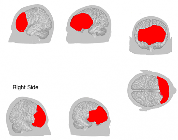
The red corresponds to patient PR’s prefrontal resection. (Credit: Gilles Lafargue)
It is important to note that patients, in this study, were patients with a diffuse low-grade glioma. Contrary to acute lesions as strokes, functional disturbances associated with this kind of tumor are generally limited, despite huge lesions and resections. Such a high degree of plasticity is explained by the tumour’s slow growth, which enables progressive, functional reorganization in a much more efficient manner than an acute lesion would.
For example, in patients with low-grade glioma, it has been possible to resect the inferior parietal lobe (Lafargue & Duffau, 2008) or the Broca’s area (Plaza, Gatignol, Leroy, & Duffau, 2009) with preserved functions traditionally thought to be relying on these damaged neural systems, that is to say awareness of intending to act and language production. Hence, the new study by Anne-Laure Lemaitre, Guillaume Herbet, Hugues Duffau and Gilles Lafargue extends to the highest level of human neurocognitive organization the ability of the brain to resist to very extensive and irreversible destruction of the neural tissue.
Such results go against the conventional idea whereby mental functions are directly and permanently linked to particular focal cortical regions. Indeed, higher-order cognitive functions are subserved by numerous subnetworks and built-in redundancy rather than by specialized structures. This yields great flexibility and cognitive resilience even in the face of the destruction of some cortical epicentres.
These findings are described in the article entitled Preserved metacognitive ability despite unilateral or bilateral anterior prefrontal resection, recently published in the journal Brain and Cognition. This work was led by Anne-Laure Lemaitre from the Hôpital Gui de Chauliac and the University of Lille, in collaboration with Guillaume Herbet and Hugues Duffau from the Hôpital Gui de Chauliac and INSERM, and Gilles Lafargue from Reims Champagne-Ardenne University.
References:
- Lafargue, G., & Duffau, H. (2008). Awareness of intending to act following parietal cortex resection. Neuropsychologia, 46(11), 2662–2667.
- Plaza, M., Gatignol, P., Leroy, M., & Duffau, H. (2009). Speaking without Broca’s area after tumor resection. Neurocase, 15(4), 294–310.
Related Posts
 Genes Keep You On A Long Leash With Your Dog
 Looking At Versus Focusing On Faces: What Attracts Our Attention?
 What Do They Get Out Of It? The Emotional Experience Of Providing Social Support
 How Does Rewarding Safe Choices Affect Teen Decision Making In Peer Contexts?
 Adolescent Self-Efficacy Is Shaped By Family, School, And Peers
 Does Morality Make Us Make God In Our Own Image?